 Indietro
Smooth Muscle Anatomy and Contraction: Structure, Types, and Comparison with Skeletal Muscle
Guida di studio - Note intelligenti
 Appunti personalizzati basati sui tuoi materiali, ampliati con definizioni chiave, esempi e contesto.
Smooth Muscle Anatomy
Overview and Location
Smooth muscle is a type of involuntary muscle tissue found in the walls of most hollow organs throughout the body. Its primary function is to facilitate the movement of substances through these organs by contracting and relaxing in a coordinated manner.
Locations: Digestive tract, respiratory tract, urinary system, cardiovascular system, reproductive system, eyes, and skin.
Organization: Most smooth muscle is organized into sheets of tightly packed fibers.
Microscopic Anatomy of Smooth Muscle
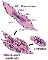
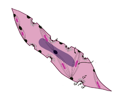
Smooth muscle cells are spindle-shaped, have a single central nucleus, and lack the striations seen in skeletal muscle. They contain actin and myosin filaments arranged diagonally, which allows for a unique corkscrew contraction.
Cell Structure: Short, tapered cells with a single nucleus.
Connective Tissue: Each cell is surrounded by a thin layer of connective tissue called the endomysium.
Ultrastructure: Poorly developed sarcoplasmic reticulum and presence of caveolae (small invaginations of the plasma membrane).
Organization of Smooth Muscle Layers
Longitudinal and Circular Layers
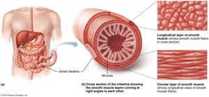
Most organs containing smooth muscle have two layers of fibers oriented at right angles to each other:
Longitudinal Layer: Fibers run parallel to the long axis of the organ; contraction shortens the organ.
Circular Layer: Fibers run around the circumference of the organ; contraction constricts the organ's lumen.
Function: Alternating contractions of these layers mix and propel substances through hollow organs (peristalsis).
Types of Smooth Muscle
Unitary (Visceral) Smooth Muscle
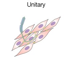
Unitary smooth muscle is the most common type, found in the walls of hollow organs. Cells are connected by gap junctions, allowing coordinated contraction as a single unit.
Structure: Cells connected by gap junctions; always contains longitudinal and circular fibers.
Control: Only a few cells receive direct nervous stimulation; the signal spreads to neighboring cells.
Location: Digestive tract, urinary bladder, uterus, small blood vessels.
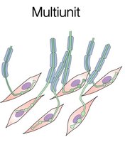
Multiunit Smooth Muscle
Multiunit smooth muscle consists of independent cells, each receiving its own nerve ending, allowing for precise control. This type is less common and not always organized in layers.
Structure: Few or no gap junctions; each cell operates independently.
Control: Each cell receives direct nervous stimulation.
Location: Large blood vessels, iris muscles of the eye, arrector pili muscles of the skin.
Comparison: Smooth vs. Skeletal Muscle
Structural Differences
Skeletal Muscle: Long, striated cells; multiple nuclei; contains myofibrils and three layers of connective tissue (epimysium, perimysium, endomysium).
Smooth Muscle: Short, tapered cells; single nucleus; no myofibrils; only endomysium as connective tissue.
Cellular Structures
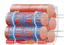
Skeletal Muscle: Well-developed sarcoplasmic reticulum and T-tubules for rapid signal transmission.
Smooth Muscle: Poorly developed sarcoplasmic reticulum; contains caveolae instead of T-tubules for calcium entry.
Arrangement of Contractile Proteins
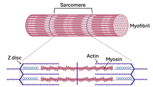
Skeletal Muscle: Contains sarcomeres with actin and myosin organized in a regular, repeating pattern anchored to Z-discs.
Smooth Muscle: Actin and myosin are arranged in diagonal chains anchored at dense bodies, resulting in a corkscrew contraction.
Smooth Muscle Contraction
Mechanism of Contraction
Smooth muscle contraction is similar to skeletal muscle in that myosin heads pull on actin filaments. However, smooth muscle can maintain contraction for longer periods with less energy due to the 'latch state,' where myosin heads remain attached to actin without continuous ATP consumption.
Latch State: Allows smooth muscle to sustain force with minimal energy expenditure.
Calcium Entry: Calcium ions enter through caveolae, triggering contraction.
Comparison Table: Skeletal vs. Smooth Muscle
Feature | Skeletal Muscle | Smooth Muscle |
|---|---|---|
Control | Voluntary | Involuntary |
Sarcolemma Indentations | T-tubules | Caveolae |
Myofilaments | Actin & Myosin | Actin & Myosin |
Contraction Speed & Strength | Rapid, powerful, fatigue easily | Slow, less powerful, resistant to fatigue (latch state) |
Summary
Smooth muscle is essential for the function of hollow organs, enabling involuntary movements such as peristalsis and vasoconstriction. Its unique structure and contraction mechanism allow for sustained activity with minimal energy use, distinguishing it from skeletal muscle in both form and function.
