 Indietro
Somatic Nervous System and Skeletal Muscle: Structure, Function, and Contraction
Guida di studio - Note intelligenti
 Appunti personalizzati basati sui tuoi materiali, ampliati con definizioni chiave, esempi e contesto.
Somatic Motor Division
Overview of Somatic Motor Pathways
The somatic motor division is responsible for voluntary control of skeletal muscles. It consists of a single neuron pathway originating in the central nervous system (CNS), specifically in the brain or the ventral horn of the spinal cord.
Single neuron pathway: The neuron is myelinated, very long, and always excitatory.
Neuromuscular junction (NMJ): The synapse between the somatic motor neuron and the skeletal muscle fiber.
Motor end plate: Specialized region of the muscle cell membrane where the neuron terminates.
Acetylcholinesterase (AChE): Enzyme in the synaptic cleft that breaks down acetylcholine.
Types of Muscle Tissue
Classification and Characteristics
Muscle tissue is classified into three types, each with distinct structural and functional properties.
Skeletal muscle: Striated, attached to bones, voluntary control, responds to somatic motor neurons.
Cardiac muscle: Striated, found only in the heart, involuntary control, moves blood, modulated by autonomic and endocrine systems.
Smooth muscle: Non-striated, found in internal organs and tubes, involuntary control, moves materials within the body, modulated by autonomic and endocrine systems.
Skeletal Muscle Structure
Organization of Skeletal Muscle
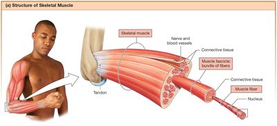
Skeletal muscle is composed of bundles of muscle fibers (cells) organized into fascicles, surrounded by connective tissue, and attached to bone via tendons.
Muscle fibers: Fused cells with multiple nuclei.
Fascicles: Bundles of muscle fibers surrounded by connective tissue.
Connective tissue: Surrounds the entire muscle and is continuous with tendons.
Muscle Fiber Anatomy
Muscle fibers have specialized terminology for their cellular structures.
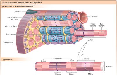
Sarcolemma: Cell membrane of the muscle fiber.
Sarcoplasm: Cytoplasm of the muscle fiber.
Sarcoplasmic reticulum: Modified endoplasmic reticulum, stores and releases Ca2+.
Transverse tubules (T-tubules): Extensions of the sarcolemma that penetrate the fiber, allowing action potentials to reach internal structures.
Triad: T-tubule flanked by two terminal cisternae of the sarcoplasmic reticulum.
Myofibrils: Contractile structures within the muscle fiber.
Mitochondria and glycogen granules: Provide energy for contraction.
T-Tubules and Sarcoplasmic Reticulum
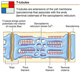
T-tubules are critical for transmitting action potentials deep into the muscle fiber, triggering Ca2+ release from the sarcoplasmic reticulum.
Triad: Structure formed by a T-tubule and two terminal cisternae.
Function: Brings action potentials to the interior, facilitating rapid contraction.
Muscle Filament Proteins
Thick and Thin Filaments
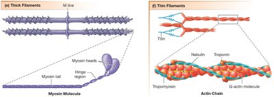
Muscle contraction is driven by the interaction of thick and thin filaments within the myofibrils.
Thick filaments: Composed primarily of myosin molecules, with heads that bind to actin.
Thin filaments: Composed of actin, troponin, tropomyosin, and associated proteins like titin and nebulin.
Sarcomere Structure
Organization and Function of the Sarcomere
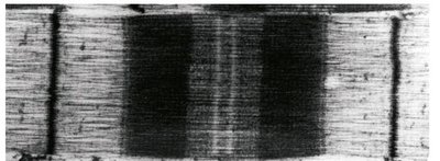
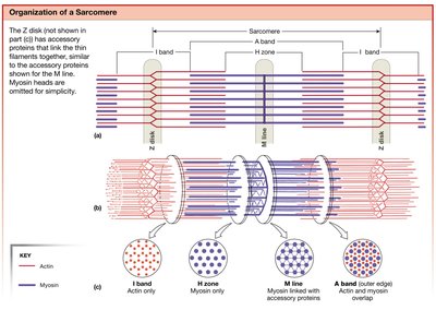
The sarcomere is the fundamental contractile unit of muscle, defined by the arrangement of thick and thin filaments.
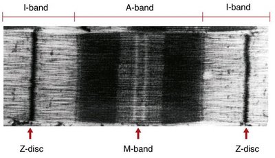
Z disks: Boundaries of the sarcomere, anchor thin filaments.
I band: Region containing only thin filaments (actin).
A band: Region where thick (myosin) and thin (actin) filaments overlap.
H zone: Central region of the A band with only thick filaments.
M line: Proteins that anchor thick filaments in the center of the sarcomere.
Sarcomere Shortening During Contraction
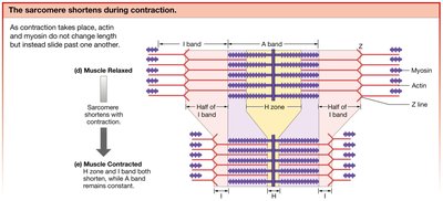
During muscle contraction, the sarcomere shortens as actin and myosin filaments slide past each other, but their lengths do not change.
Sliding filament theory: Explains how muscle contraction occurs by the sliding of filaments.
H zone and I band: Both shorten during contraction, while the A band remains constant.
Muscle Fiber Concept Map
Hierarchical Organization of Skeletal Muscle
The concept map illustrates the structural hierarchy from connective tissue to sarcomere, highlighting the relationships between components. 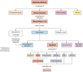
Summary of Muscle Contraction
Key Steps in Muscle Contraction
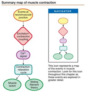
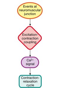
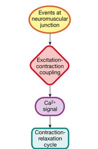
Muscle contraction involves a sequence of events starting at the neuromuscular junction, followed by excitation-contraction coupling, Ca2+ signaling, and the contraction-relaxation cycle.
Muscle twitch: Single contraction-relaxation event.
Sliding filament theory: Mechanism of contraction.
Neuromuscular Junction
Structure and Function
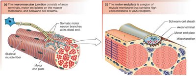
The neuromuscular junction is the site where the somatic motor neuron communicates with the skeletal muscle fiber.
Axon terminals: Release acetylcholine (ACh) into the synaptic cleft.
Motor end plate: Region of muscle membrane with high concentration of ACh receptors.
Events at the Motor End Plate
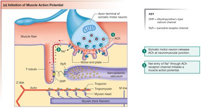
The release of ACh and its binding to receptors initiates muscle action potentials.
ACh release: Triggered by action potential arriving at the axon terminal.
Binding to nicotinic receptors: Opens ion channels, allowing Na+ influx and depolarization.
Excitation-Contraction Coupling
Initiation of Muscle Action Potential
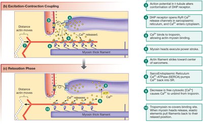
Excitation-contraction coupling links the electrical signal from the neuron to the mechanical contraction of the muscle.
Action potential: Travels along the sarcolemma and T-tubules.
Ca2+ release: From the sarcoplasmic reticulum, triggered by DHP and RyR channels.
Contraction: Ca2+ binds to troponin, allowing myosin to bind actin.
Excitation-Contraction Coupling and Relaxation
Coupling: Action potential in T-tubule alters DHP receptor, opening RyR Ca2+ channels.
Relaxation: Ca2+ is pumped back into the sarcoplasmic reticulum by SERCA ATPase, and troponin-tropomyosin complex returns to resting state.
Troponin and Tropomyosin
Regulation of Contraction
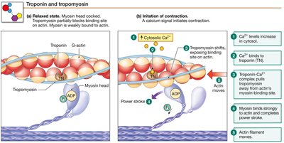
Troponin and tropomyosin regulate the binding of myosin to actin, controlling muscle contraction.
Resting state: Tropomyosin blocks myosin-binding sites on actin.
Contraction: Ca2+ binds to troponin, shifting tropomyosin and exposing binding sites.
Contraction Cycle
Steps of the Contraction Cycle
The contraction cycle describes the sequence of events that allow myosin and actin to interact, resulting in muscle contraction.
ATP binding: Releases myosin from actin.
ATP hydrolysis: Energizes myosin head.
Cross-bridge formation: Myosin binds actin.
Power stroke: Myosin head pivots, pulling actin filament.
ADP release: Myosin remains bound until ATP binds again.
Speed of Muscle Twitch
Phases of Muscle Twitch
Muscle twitch consists of three phases: latent period, contraction, and relaxation.
Latent period: Excitation-contraction coupling; no tension.
Contraction: Cross-bridge formation; tension increases.
Relaxation: Ca2+ reentry into SR; tension declines.
Graded Muscle Responses
Mechanisms of Graded Contraction
Muscle contraction strength is graded by frequency of stimulation and recruitment of motor units.
Temporal summation: Increased stimulus frequency leads to greater force.
Motor unit recruitment: More motor units activated for stronger contractions.
Motor Units and Recruitment
Size Principle and Force Production
Motor units are recruited based on size, from smallest to largest, to control contraction force.
Small motor units: Activated first, produce small force.
Large motor units: Activated last, produce large force.
Muscle Fiber Types
Comparison of Muscle Fiber Types
Muscle fibers are classified based on their contraction speed, metabolism, and fatigue resistance.
Type | Speed | Myosin ATPase Activity | Diameter | Duration | SR ATPase Activity | Endurance | Use | Metabolism | Capillary Density | Mitochondria | Color |
|---|---|---|---|---|---|---|---|---|---|---|---|
Slow-Twitch Oxidative (Type I) | Slowest | Slow | Small | Longest | Moderate | Fatigue resistant | Posture | Oxidative; aerobic | High | Numerous | Dark red |
Fast-Twitch Oxidative-Glycolytic (Type IIA) | Intermediate | Fast | Medium | Short | High | Fatigue resistant | Standing, walking | Glycolytic, more oxidative with training | Medium | Moderate | Red |
Fast-Twitch Glycolytic (Type IIB/IIX) | Fastest | Fast | Large | Short | High | Easily fatigued | Jumping, quick movements | Glycolytic; anaerobic | Low | Few | Pale |
Length-Tension Relationships
Optimal Sarcomere Length for Force Production
The amount of tension a muscle can produce depends on the initial length of the sarcomere.
Optimal length: Maximum overlap of actin and myosin allows greatest force.
Too short or too long: Reduced overlap decreases force production.
