 Indietro
Special Senses: Olfaction, Gustation, Vision, and Hearing
Guida di studio - Note intelligenti
 Appunti personalizzati basati sui tuoi materiali, ampliati con definizioni chiave, esempi e contesto.
Special Senses
Overview of Special Senses
The special senses include olfaction (smell), gustation (taste), vision, equilibrium (balance), and hearing. These senses rely on specialized organs and neural pathways to detect and interpret environmental stimuli.
Olfaction: Sense of smell, mediated by olfactory organs in the nasal cavity.
Gustation: Sense of taste, mediated by taste buds on the tongue and other oral structures.
Vision: Sense of sight, mediated by the eyes and associated structures.
Equilibrium: Sense of balance, mediated by the vestibular system in the inner ear.
Hearing: Sense of sound, mediated by the auditory system in the ear.
Olfaction (Sense of Smell)
Anatomy of Olfactory Organs
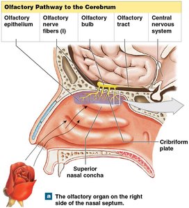
Olfactory organs are located in the nasal cavity, on either side of the nasal septum. They consist of two main layers: the olfactory epithelium and the lamina propria.
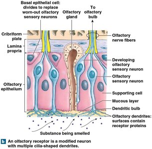
Olfactory epithelium: Contains olfactory sensory neurons, supporting cells, and basal epithelial cells (stem cells).
Lamina propria: Contains areolar tissue, blood vessels, nerves, and olfactory glands that produce mucus.
Olfactory Reception and Pathways
Olfactory reception begins when odorant molecules bind to G protein-coupled receptors on olfactory sensory neurons, generating a depolarization (generator potential).
Pathway: Afferent fibers from olfactory neurons collect into bundles, penetrate the cribriform plate, and synapse in the olfactory bulbs. Axons from the bulbs travel along the olfactory tract to the olfactory cortex, hypothalamus, and limbic system.
Direct cortical access: Olfactory information is unique in reaching the cerebral cortex directly, without thalamic relay.
Olfactory Discrimination and Adaptation
Humans can distinguish thousands of odors, though the number of olfactory neurons declines with age. Dogs have a much greater olfactory receptor surface area, making their sense of smell far superior.
Olfactory receptor renewal: Olfactory receptors are replaced frequently by basal epithelial cells.
Discrimination: Ability to detect 2000–4000 odors.
Gustation (Sense of Taste)
Anatomy of Taste Buds and Lingual Papillae
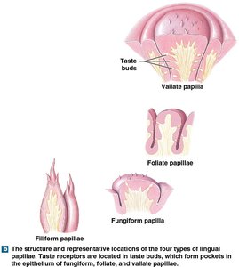
Gustatory epithelial cells (taste receptors) are found in taste buds, which are distributed on the superior surface of the tongue and parts of the pharynx and larynx. Taste buds are associated with lingual papillae.
Types of lingual papillae:
Filiform papillae: Provide friction, do not contain taste buds.
Fungiform papillae: Contain about five taste buds each.
Vallate papillae: Contain up to 100 taste buds each.
Foliate papillae: Have taste buds.
Gustatory Discrimination
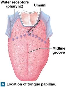
Humans perceive six basic taste sensations: sweet, salty, sour, bitter, umami, and water.
Primary tastes: Sweet, salty, sour, bitter.
Umami: Pleasant, savory taste associated with glutamate.
Water: Detected by water receptors in the pharynx.
Vision (Sense of Sight)
Accessory Structures of the Eye
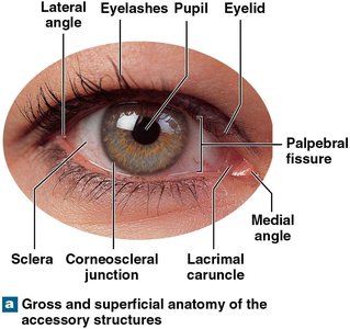
The eye is protected and supported by accessory structures, including the eyelids, superficial epithelium, and lacrimal apparatus.
Eyelids: Protect the eye and help distribute tears.
Lacrimal apparatus: Produces and drains tears.
Superficial epithelium: Covers the exposed surface of the eye.
Sectional Anatomy of the Eye
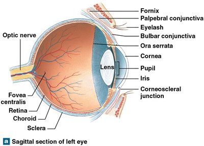
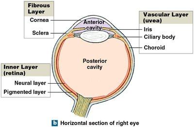
The eye consists of three main layers: fibrous, vascular, and inner (retina). Each layer has distinct functions and structures.
Fibrous layer: Includes the cornea and sclera.
Vascular layer (uvea): Includes the iris, ciliary body, and choroid.
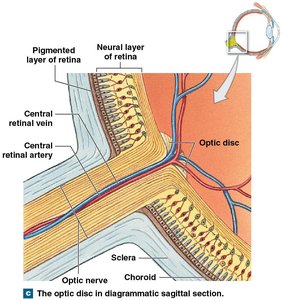
Inner layer (retina): Includes the pigmented layer and neural layer.
Pupillary Muscles
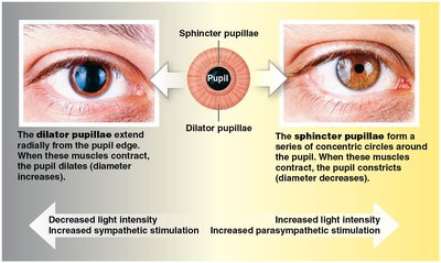
The iris contains two types of smooth muscle: the dilator pupillae and sphincter pupillae, which control the diameter of the pupil in response to light intensity.
Dilator pupillae: Radially arranged, dilate the pupil under sympathetic stimulation.
Sphincter pupillae: Circularly arranged, constrict the pupil under parasympathetic stimulation.
Retina and Photoreceptors
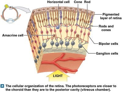
The retina is the innermost layer of the eye, responsible for converting light into neural signals. It contains two types of photoreceptors: rods and cones.
Rods: Highly sensitive to light, do not discriminate colors.
Cones: Provide color vision, concentrated in the macula and fovea centralis.
Visual axis: Line from an object to the fovea, site of sharpest vision.
Hearing and Equilibrium (Auditory System)
Anatomy of the Ear
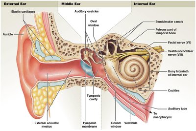
The ear is divided into three regions: external, middle, and internal ear. Each region has specialized structures for hearing and balance.
External ear: Includes the auricle, external acoustic meatus, tympanic membrane, and ceruminous glands.
Middle ear: Contains the tympanic cavity, auditory tube, and three auditory ossicles (malleus, incus, stapes).
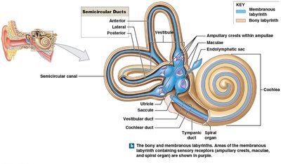
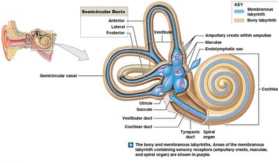
Internal ear: Composed of the bony and membranous labyrinth, including the vestibule, semicircular canals, and cochlea.
Middle Ear Structures and Function
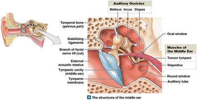
The middle ear transmits sound vibrations from the tympanic membrane to the internal ear via the auditory ossicles. Two small muscles (tensor tympani and stapedius) protect the ear from loud noises.
Auditory ossicles: Malleus, incus, stapes.
Tensor tympani: Stiffens tympanic membrane.
Stapedius: Reduces movement of stapes at oval window.
Internal Ear: Hearing and Equilibrium
The internal ear contains the vestibule, semicircular canals, and cochlea. The vestibule detects gravity and linear acceleration, semicircular canals detect rotational movement, and the cochlea is responsible for hearing.
Vestibule: Contains saccule and utricle.
Semicircular canals: Contain semicircular ducts.
Cochlea: Contains cochlear duct and sensory receptors for hearing.
Process of Hearing
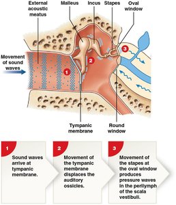
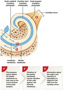
Hearing involves six basic steps, from the arrival of sound waves to the transmission of neural signals to the brain.
Sound waves arrive at the tympanic membrane.
Movement of the tympanic membrane displaces the auditory ossicles.
Movement of the stapes at the oval window produces pressure waves in the perilymph of the scala vestibuli.
The pressure waves distort the basilar membrane on their way to the round window of the scala tympani.
Vibration of the basilar membrane causes hair cells to vibrate against the tectorial membrane.
Information about the region and intensity of stimulation is relayed to the CNS over the cochlear nerve.
Summary Table: Special Senses and Their Organs
Sense | Organ | Main Receptors | Pathway |
|---|---|---|---|
Olfaction | Nasal cavity | Olfactory sensory neurons | Olfactory bulb → Olfactory tract → Cortex |
Gustation | Tongue, pharynx, larynx | Gustatory epithelial cells | Taste buds → Cranial nerves → Brainstem → Cortex |
Vision | Eye | Rods and cones | Retina → Optic nerve → Thalamus → Cortex |
Hearing | Ear (cochlea) | Hair cells | Cochlear nerve → Brainstem → Cortex |
Equilibrium | Ear (vestibule, semicircular canals) | Hair cells | Vestibular nerve → Brainstem → Cortex |
Additional info: Academic context was added to clarify neural pathways, receptor types, and the functional significance of each special sense.
