 Indietro
Special Senses: Vision – Anatomy and Physiology of the Eye
Guida di studio - Note intelligenti
 Appunti personalizzati basati sui tuoi materiali, ampliati con definizioni chiave, esempi e contesto.
Special Senses: Vision
Anatomy of the Eye
The human eye is a complex organ responsible for detecting light and converting it into neural signals for vision. Its structure is organized into several distinct layers and compartments, each with specialized functions.
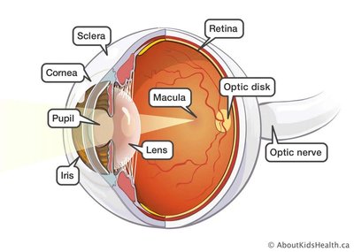
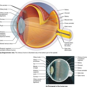
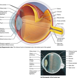
Fibrous Layer: The outermost layer, consisting of the cornea and sclera. The cornea is transparent and avascular, allowing light to enter, while the sclera provides structural support.
Vascular Layer: Includes the choroid, ciliary body, and iris. The choroid is pigmented and nourishes the eye, the ciliary body controls lens shape and produces aqueous humor, and the iris regulates pupil size.
Neural Layer: The retina contains photoreceptors (rods and cones) that detect light and initiate visual signals.
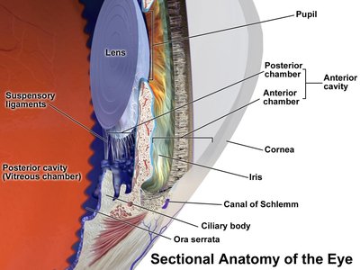
Anterior and Posterior Segments: The eye is divided into anterior and posterior segments, separated by the lens. The anterior segment contains aqueous humor, while the posterior segment contains vitreous humor.
Surface Anatomy and External Structures
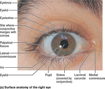
The external anatomy of the eye includes protective and lubricating structures that maintain ocular health and facilitate vision.
Eyelids and Eyelashes: Protect the eye from debris and injury.
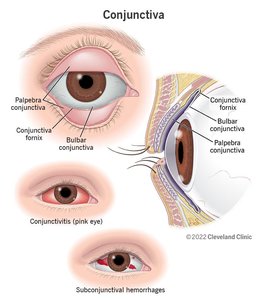
Conjunctiva: A thin membrane covering the sclera and lining the eyelids, providing lubrication and protection.
Lacrimal Gland: Produces tears to lubricate the eye and remove irritants.
Palpebral and Bulbar Conjunctiva: Different regions of the conjunctiva, with the palpebral covering the inside of the eyelids and the bulbar covering the sclera.
Eye Cavities and Chambers
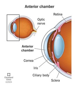
The eye contains two main cavities: the anterior cavity (filled with aqueous humor) and the posterior cavity (filled with vitreous humor). These fluids maintain intraocular pressure and provide nutrients.
Anterior Chamber: Located between the cornea and iris, filled with aqueous humor.
Posterior Chamber: Located between the iris and lens, also filled with aqueous humor.
Vitreous Chamber: Located behind the lens, filled with vitreous humor, which helps maintain the shape of the eye and holds the retina in place.
Fibrous Layer: Cornea and Sclera
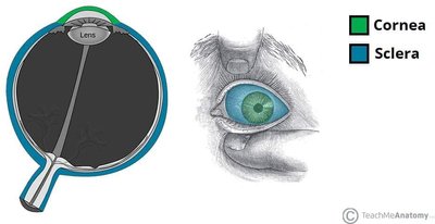
The fibrous layer provides protection and structure. The cornea is transparent and allows light to enter, while the sclera is opaque and forms the white of the eye.
Cornea: Avascular, transparent, and responsible for most of the eye's refractive power.
Sclera: Dense connective tissue that maintains the shape of the eyeball and provides attachment for extraocular muscles.
Vascular Layer: Choroid, Ciliary Body, and Iris
The vascular layer is essential for nourishing the eye and regulating the amount of light entering the eye.
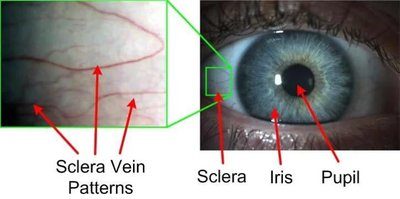
Choroid: Contains blood vessels and pigment to absorb excess light.
Ciliary Body: Contains muscles that adjust the shape of the lens for focusing and produces aqueous humor.
Iris: Contains muscles that control the size of the pupil, regulating light entry.
Pupillary Muscles: Sphincter pupillae (parasympathetic, constricts pupil) and dilator pupillae (sympathetic, dilates pupil).
Corneal Donation and Immune Privilege
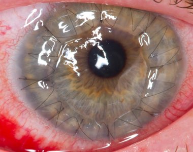
The cornea's avascular nature makes it suitable for transplantation, as immune responses are less aggressive in the eye. Blood-type matching is usually unnecessary due to the eye's immune privilege.
Corneal Transplant: Surgical procedure to replace a damaged or diseased cornea.
Immune Privilege: Reduced immune response in the eye, allowing for easier transplantation.
Extraocular Muscles and Cranial Nerves
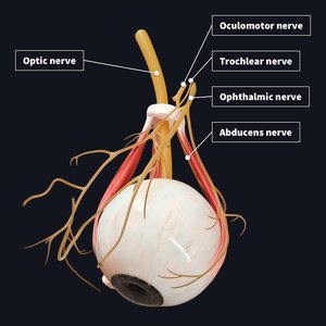
Movement of the eye is controlled by six extraocular muscles, which are innervated by three cranial nerves: oculomotor (III), trochlear (IV), and abducens (VI). The optic nerve (II) transmits visual information to the brain.
Oculomotor Nerve (III): Controls most extraocular muscles.
Trochlear Nerve (IV): Controls the superior oblique muscle.
Abducens Nerve (VI): Controls the lateral rectus muscle.
Optic Nerve (II): Transmits visual signals from the retina to the brain.
Dynamics of Vision
Light Transmission and Refraction
Light enters the eye and is refracted (bent) as it passes through the cornea, aqueous humor, lens, and vitreous humor. The lens focuses light onto the macula lutea, the area of highest visual acuity.
Refraction: Light is bent three times: entering the cornea, entering the lens, and leaving the lens.
Macula Lutea: Central, cone-rich area of the retina specialized for sharp, detailed, color vision.
Accommodation: Focusing on Near and Distant Objects
Accommodation is the process by which the eye adjusts its focus for near or distant objects. This involves changes in the shape of the lens, controlled by the ciliary muscles and zonular fibers.
Distant Vision: Ciliary muscles relaxed, zonular fibers taut, lens is flat.
Near Vision: Ciliary muscles contract, zonular fibers loosen, lens becomes round.
Retina and Photoreceptors
The retina contains two types of photoreceptors: rods (for low-light and peripheral vision) and cones (for color and high-acuity vision). Photoreceptors are embedded in the pigmented layer and contain visual pigments that initiate the signal to light.
Rods: Sensitive to dim light, provide black-and-white vision.
Cones: Sensitive to bright light, provide color vision and high visual acuity.
Visual Pigments: Proteins (opsins) combined with retinal (vitamin A derivative) that change shape in response to light.
Phototransduction Cascade
Phototransduction is the process by which light is converted into electrical signals in the retina. The visual pigment rhodopsin undergoes a series of changes in response to light, triggering a G-protein cascade.
Pigment Synthesis: In the dark, 11-cis-retinal combines with opsin to form rhodopsin.
Pigment Bleaching: Light converts 11-cis-retinal to all-trans-retinal, separating it from opsin and triggering the cascade.
Pigment Regeneration: Enzymes convert all-trans-retinal back to 11-cis-retinal, reforming rhodopsin.
G-Protein Cascade: Light activates transducin, which activates phosphodiesterase (PDE), reducing cGMP levels and closing cation channels, leading to hyperpolarization of the photoreceptor.
Key Equations:
Photoreceptor hyperpolarization: (in light)
Visual Pathways and Field Perception
Visual signals are transmitted from the retina to the brain via the optic nerve. The retina is divided into nasal and temporal portions; nasal fibers cross at the optic chiasm, allowing for binocular vision and field perception.
Nasal Retina: Perceives outer visual field; fibers cross at the optic chiasm.
Temporal Retina: Perceives inner visual field; fibers do not cross.
Optic Nerve: Carries action potentials generated by photoreceptors to the brain for processing.
Visual Acuity
Visual acuity is the clarity or sharpness of vision, measured by the ability to see fine details at a set distance. For example, 20/40 vision means a person must stand 20 feet away to see what a person with normal vision can see from 40 feet away.
Summary of Key Concepts
Accommodation is used for close vision and involves the lens, pupil, and extraocular muscles.
Vision utilizes a three-neuron system starting with proteins on specialized photoreceptors.
G-protein signaling cascade is essential for phototransduction.
Color-blindness is a genetic disorder affecting photoreceptors, often X-linked recessive.
Retinas are divided into nasal and temporal portions; nasal portions cross in the optic chiasm, and different regions of damage result in different visual field deficits.
