 Indietro
Spinal Cord, Brain, and Reflexes: Structure and Function
Guida di studio - Note intelligenti
 Appunti personalizzati basati sui tuoi materiali, ampliati con definizioni chiave, esempi e contesto.
Spinal Cord: Structure and Function
Gross Anatomy of the Spinal Cord
The spinal cord is a major component of the central nervous system, housed within the vertebral column and protected by specialized membranes called meninges. It serves as a conduit for sensory and motor information between the brain and the rest of the body, and gives rise to spinal nerves.
Length: Approximately 45 cm (18 in.) long and 14 mm (1/2 in.) wide.
Regions: Divided into cervical, thoracic, lumbar, and sacral regions.
Symmetry: Exhibits bilateral symmetry.
Spinal Meninges
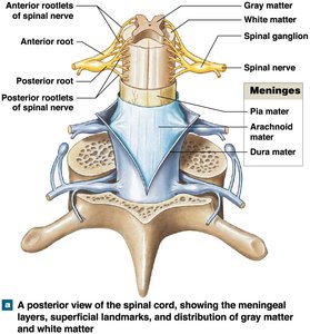
The spinal cord is surrounded by three meningeal layers that protect it and carry blood supply. These layers are continuous with the cranial meninges.
Dura mater: Outermost, tough layer.
Arachnoid mater: Middle, web-like layer.
Pia mater: Innermost, delicate layer attached to the spinal cord surface.
Function: Protection and vascular supply.
Meningitis: Inflammation due to infection (viral or bacterial).
Gray Matter and White Matter
The spinal cord contains both gray and white matter, each with distinct structural and functional roles.
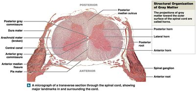
Gray matter: Composed of neuron cell bodies, neuroglia, and unmyelinated axons. Organized into nuclei and horns.
White matter: Contains myelinated and unmyelinated axons, organized into columns.
Functional and Structural Organization
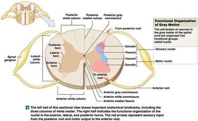
Gray matter is organized into horns, each with specific functions:
Posterior horns: Somatic and visceral sensory nuclei (incoming information).
Anterior horns: Somatic motor nuclei (outgoing information).
Lateral horns: Visceral motor nuclei (thoracic and lumbar segments).
White matter is organized into columns:
Posterior white columns: Between posterior horns and posterior median sulcus.
Anterior white columns: Between anterior horns and anterior median fissure.
Lateral white columns: On each side between anterior and posterior columns.
Anterior white commissure: Axons cross from one side to the other.
Dermatomes
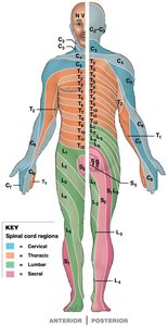
Dermatomes are regions of skin innervated by specific spinal nerves. They are important for clinical assessment of nerve function and injury.
Distribution: Each spinal segment innervates a specific region.
Overlap: Adjacent segments may overlap.
Motor vs. Sensory: Slight differences in distribution.
Spinal Reflexes
Neural Reflexes and Reflex Arc
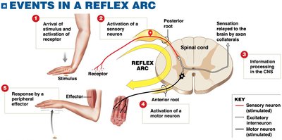
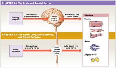
Reflexes are rapid, automatic responses to specific stimuli, forming the basic building blocks of neural function. Each reflex produces a consistent motor response.
Components of a reflex arc:
Sensory receptor
Sensory neuron
Information processing in CNS
Motor neuron
Effector (muscle/gland)
Events in a spinal reflex arc:
Stimulus activates a receptor
Action potential generated in sensory neuron (enters via posterior root)
Information processing (interneurons in spinal cord)
Motor neuron activated (axon leaves via anterior root)
Effector stimulated
Brain: Structure and Function
Overview of Brain Regions
The adult human brain contains nearly all of the body's nervous tissue and is divided into four main regions, each with distinct functions.
Cerebrum: Largest part, controls higher mental functions (conscious thought, intellect, memory).
Cerebellum: Coordinates repetitive body movements, covered by gray matter.
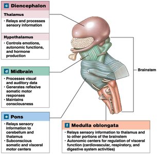
Diencephalon: Contains thalamus and hypothalamus, relays and processes sensory information, controls emotions and hormone production.
Brainstem: Relays information between spinal cord and cerebrum/cerebellum; includes midbrain, pons, and medulla oblongata.
Cerebrum
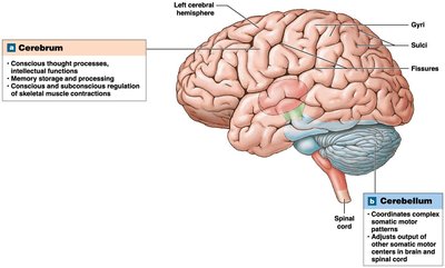
The cerebrum is the largest region of the brain, divided into left and right hemispheres. Its surface layer, the cerebral cortex, is composed of gray matter. Rounded elevations called gyri increase surface area, separated by sulci (shallow depressions) and fissures (deep grooves).
Functions: Conscious thought, intellect, memory, regulation of skeletal muscle contractions.
Cerebellum
The cerebellum is the second-largest part of the brain, responsible for coordinating complex motor patterns and repetitive movements. It consists of two hemispheres and is covered by the cerebellar cortex (gray matter).
Functions: Coordination of movement, adjustment of motor output.
Brainstem
The brainstem relays information between the spinal cord and higher brain regions. It consists of the midbrain, pons, and medulla oblongata, each with specialized functions.
Midbrain: Processes visual and auditory data, generates reflexive motor responses, maintains consciousness.
Pons: Connects cerebellum to brainstem, contains relay centers and nuclei for motor control.
Medulla oblongata: Connects brain to spinal cord, relays sensory information, regulates autonomic functions (heart rate, blood pressure, digestion).
Brain Protection and Support
The brain is protected by cranial meninges, which are continuous with the spinal meninges and consist of three layers:
Dura mater: Outer fibrous layer (periosteal cranial dura) fused to periosteum, and inner fibrous layer (meningeal cranial dura).
Arachnoid mater: Covers brain, attaches to dura mater, separated by subdural space; subarachnoid space lies between arachnoid and pia mater.
Pia mater: Attached to brain surface by astrocytes.
Summary Table: Spinal Cord and Brain Regions
Region | Main Function | Key Structures |
|---|---|---|
Spinal Cord | Relays sensory and motor information; reflexes | Gray matter (horns), white matter (columns), meninges |
Cerebrum | Conscious thought, intellect, memory | Gyri, sulci, fissures, cortex |
Cerebellum | Coordination of movement | Cerebellar cortex, hemispheres |
Diencephalon | Relay/process sensory info, hormone control | Thalamus, hypothalamus |
Brainstem | Relay info, autonomic functions | Midbrain, pons, medulla oblongata |
Key Equations and Concepts
Action Potential: The change in membrane potential that allows neurons to transmit signals.
Reflex Arc: Sequence of neural events from stimulus to response.
