 Indietro
Spinal Cord, Nerves, Reflexes, and Brain: Structure and Function
Guida di studio - Note intelligenti
 Appunti personalizzati basati sui tuoi materiali, ampliati con definizioni chiave, esempi e contesto.
Spinal Cord, Nerves, and Reflexes
Five Steps of the Reflex Arc
The reflex arc is a fundamental neural pathway that mediates a rapid, automatic response to a stimulus. It consists of five essential steps:
Stimulation of a Receptor: Detection of a stimulus by a sensory receptor.
Activation of a Sensory Neuron: Transmission of the sensory signal toward the central nervous system (CNS).
Information Processing in the CNS: Integration and processing of the sensory input, often involving interneurons.
Activation of a Motor Neuron: Transmission of the processed signal to an effector via a motor neuron.
Response of a Peripheral Effector: The effector (muscle or gland) carries out the response.
Gross Anatomy of the Spinal Cord
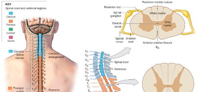
The spinal cord is a cylindrical structure approximately 45 cm long and 14 mm wide, extending from the brain to the level between the first and second lumbar vertebrae (L1–L2). It is divided into four regions and 31 segments, each associated with a pair of spinal nerves:
Cervical
Thoracic
Lumbar
Sacral
Gray matter is most prominent in segments controlling the limbs, forming the cervical and lumbosacral enlargements. The spinal cord features a posterior median sulcus (shallow groove) and an anterior median fissure (deeper groove). The central canal, filled with cerebrospinal fluid (CSF), runs through the center. Inferiorly, the conus medullaris tapers, and the filum terminale extends to the coccyx for support.
Spinal Roots and Ganglia
Each spinal segment is associated with a pair of spinal ganglia (dorsal root ganglia):
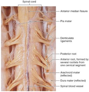
Posterior (dorsal) roots: Contain sensory neuron axons entering the spinal cord.
Anterior (ventral) roots: Contain motor neuron axons exiting the spinal cord.
Roots fan into rootlets before merging into spinal nerves, which exit via intervertebral foramina.
In adults, the lower spinal roots (L2–S5) extend beyond the spinal cord, forming the cauda equina.
Spinal Nerves and Rami
Distal to the ganglion, sensory and motor roots merge to form a mixed spinal nerve, which branches into:
Rami communicantes: White (myelinated) and gray (unmyelinated) branches serving glands and smooth muscle.
Posterior ramus: Innervates skin and muscles of the back.
Anterior ramus: Innervates ventrolateral body wall and limbs.
There are 31 pairs of mixed spinal nerves, named according to their vertebral level.
Spinal Meninges
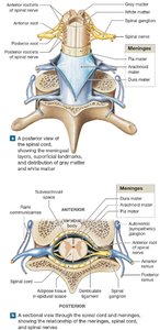
The spinal cord is protected by three connective tissue layers:
Dura mater: Outermost, tough collagenous layer; epidural space contains fat and blood vessels.
Arachnoid mater: Middle layer with a web-like structure; subarachnoid space contains CSF.
Pia mater: Innermost, delicate layer adhering to the spinal cord; denticulate ligaments anchor the cord laterally.
Functional Organization of Gray and White Matter
Gray matter consists of neuron cell bodies, neuroglia, and unmyelinated axons, organized into horns (posterior, lateral, anterior) and commissures. White matter contains myelinated and unmyelinated axons, organized into columns (posterior, lateral, anterior) and tracts:
Ascending tracts: Carry sensory information to the brain.
Descending tracts: Carry motor commands from the brain.
Anatomy of Spinal Nerves
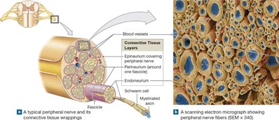
Spinal nerves are surrounded by connective tissue layers:
Epineurium: Outermost, dense collagen fibers.
Perineurium: Middle, divides nerve into fascicles.
Endoneurium: Innermost, surrounds individual axons.
Dermatomes and Peripheral Nerve Distribution
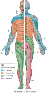
A dermatome is a skin region innervated by a single pair of spinal nerves. Loss of sensation in a dermatome can indicate nerve or spinal cord injury. Peripheral neuropathies are regional losses of sensory/motor function due to nerve trauma or compression.
Nerve Plexuses
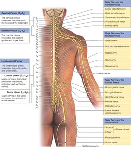
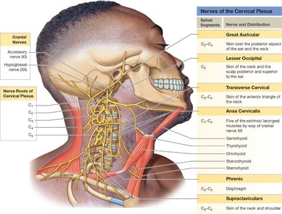
Nerve plexuses are networks formed by the anterior rami of spinal nerves, providing compound innervation to muscles and skin. The four major plexuses are:
Cervical plexus (C1–C5): Innervates neck and diaphragm (phrenic nerve).
Brachial plexus (C5–T1): Innervates pectoral girdle and upper limb.
Lumbar plexus (T12–L4): Innervates pelvic girdle and lower limb.
Sacral plexus (L4–S4): Innervates pelvic girdle and lower limb (sciatic nerve).
Neuronal Pools and Neural Circuits
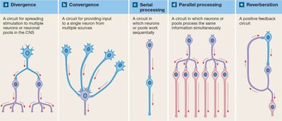
Neuronal pools are groups of interconnected interneurons with specific functions. Five basic neural circuit patterns exist:
Divergence: One neuron sends signals to multiple targets.
Convergence: Multiple neurons synapse on a single neuron.
Serial processing: Neurons work in sequence.
Parallel processing: Several neurons process the same information simultaneously.
Reverberation: Positive feedback loop maintains activity.
Neural Reflexes
Reflexes are rapid, automatic responses to stimuli, typically negative feedback loops. Reflexes can be classified by:
Development: Innate (genetically programmed) or acquired (learned).
Response: Somatic (skeletal muscle) or visceral (autonomic).
Circuit complexity: Monosynaptic (single synapse) or polysynaptic (multiple synapses).
Site of processing: Spinal or cranial.
Monosynaptic vs. Polysynaptic Reflexes
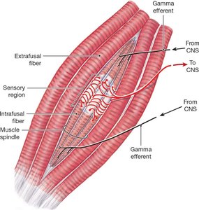
Monosynaptic reflexes: Fastest, involve a single synapse (e.g., stretch reflex, knee-jerk). Muscle spindles are the sensory receptors involved.
Polysynaptic reflexes: Involve interneurons, multiple synapses, and more complex responses (e.g., withdrawal reflex, tendon reflex, crossed extensor reflex).
The Brain and Cranial Nerves
Major Regions of the Brain
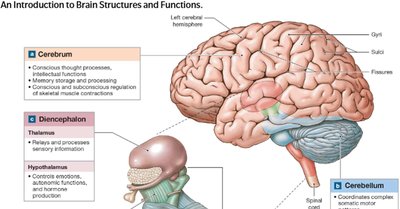
The brain is divided into four main regions:
Cerebrum: Conscious thought, memory, intellect, and voluntary movement.
Cerebellum: Coordination of movement and balance.
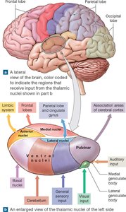
Diencephalon: Thalamus (sensory relay), hypothalamus (autonomic and endocrine control).
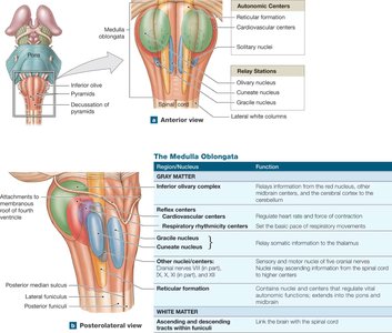
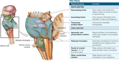
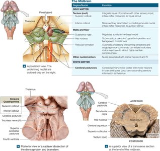
Brainstem: Midbrain, pons, and medulla oblongata (autonomic and reflex centers).
Embryology of the Brain
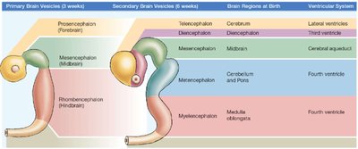
The CNS develops from the neural tube, forming three primary brain vesicles (prosencephalon, mesencephalon, rhombencephalon), which further subdivide into adult brain regions and ventricles.
Ventricles of the Brain
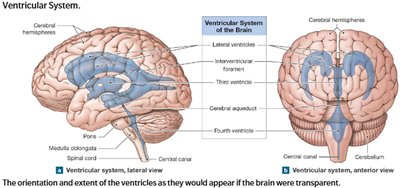
The brain contains four ventricles (two lateral, third, and fourth) filled with CSF, connected by passageways (interventricular foramen, cerebral aqueduct). Ependymal cells lining the ventricles produce and circulate CSF.
Cranial Meninges, CSF, and Blood-Brain Barrier
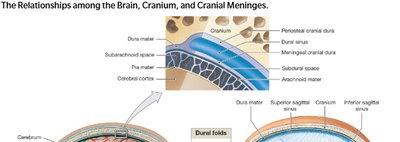
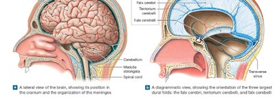
The brain is protected by three meninges (dura mater, arachnoid mater, pia mater), continuous with the spinal meninges. The dura mater forms dural folds and venous sinuses for stabilization and venous drainage. The blood-brain barrier restricts passage of substances into the CNS, maintained by tight junctions and astrocytes.
The Brainstem: Medulla Oblongata, Pons, and Midbrain
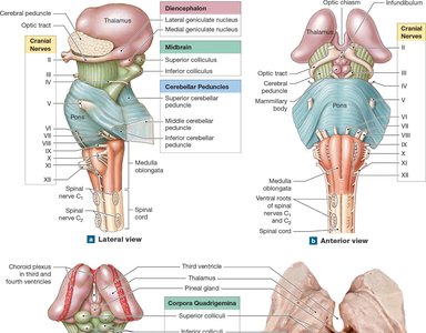
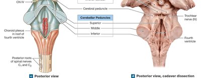
Medulla oblongata: Relays sensory and motor information, contains autonomic centers for cardiovascular and respiratory regulation.
Pons: Connects cerebellum to brainstem, contains nuclei for respiration and cranial nerves.
Midbrain: Processes visual and auditory data, maintains consciousness, contains red nucleus and substantia nigra.
The Diencephalon: Thalamus and Hypothalamus
The thalamus acts as a sensory relay and filter, while the hypothalamus regulates autonomic functions, hormone secretion, body temperature, and circadian rhythms.
The Limbic System
The limbic system is a functional network involved in emotion, motivation, and memory. It includes the cingulate gyrus, hippocampus, amygdala, and related structures.
The Cerebrum: Structure and Function
The cerebrum is divided into lobes (frontal, parietal, temporal, occipital, insula) and contains gyri and sulci to increase surface area. White matter consists of association, commissural, and projection fibers. Basal nuclei coordinate movement patterns and muscle tone.
Motor, Sensory, and Association Areas
Primary motor cortex: Controls voluntary movements.
Primary somatosensory cortex: Receives sensory input from the body.
Association areas: Interpret sensory input and coordinate motor output.
Integrative Centers and Higher Mental Functions
Integrative centers (e.g., prefrontal cortex, Wernicke's area, Broca's area) coordinate complex processes such as language, reasoning, and personality. Hemispheric lateralization refers to the specialization of each cerebral hemisphere for certain functions.
Cranial Nerves
There are 12 pairs of cranial nerves, each with specific sensory, motor, or mixed functions. They are numbered I–XII and named according to their primary function or distribution.
Sensory Pathways and Receptors
Sensory and Motor Pathways
Somatic and visceral sensory pathways carry information to the CNS, while motor pathways carry commands to effectors. Sensory receptors detect stimuli and transduce them into action potentials.
General Receptor Classification
Nociceptors: Pain receptors, tonic, large receptive fields.
Thermoreceptors: Detect temperature changes, phasic, adapt quickly.
Mechanoreceptors: Detect physical distortion (touch, pressure, vibration, proprioception).
Chemoreceptors: Detect chemical changes in body fluids.
Somatic Sensory Pathways
Spinothalamic pathway: Crude touch, pain, temperature.
Posterior column pathway: Fine touch, vibration, proprioception.
Spinocerebellar pathway: Proprioceptive information to cerebellum (unconscious).
Somatic Motor Pathways
Corticospinal (pyramidal) pathway: Voluntary control of skeletal muscles.
Medial and lateral pathways: Control of muscle tone, posture, and gross/fine movements.
Basal nuclei and cerebellum: Modulate and coordinate motor activity.
Additional info: This guide integrates and expands upon the provided material, ensuring a comprehensive, exam-ready overview of spinal cord, nerve, reflex, and brain anatomy and physiology, with direct references to the most relevant images for each topic.
