 Indietro
Step-by-Step Guidance for Nervous System Practice Test (Anatomy & Physiology)
Guida di studio - Note intelligenti
 Appunti personalizzati basati sui tuoi materiali, ampliati con definizioni chiave, esempi e contesto.
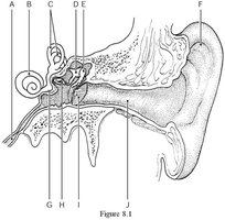
Q287. Using Figure 8.1, identify the auricle (pinna).
Background
Topic: Anatomy of the Ear
This question tests your ability to recognize the external structures of the ear, specifically the auricle (pinna), which is the visible part of the ear that collects sound waves.
Key Terms:
Auricle (pinna): The external, cartilaginous part of the ear.
External ear: Includes the auricle and external acoustic meatus.
Step-by-Step Guidance
Examine Figure 8.1 and locate the structure that forms the outermost visible part of the ear.
Identify which label points to the cartilaginous, funnel-shaped structure.
Compare the labels to the anatomical position of the auricle (pinna).
Try solving on your own before revealing the answer!
Final Answer: Label F
The auricle (pinna) is indicated by Label F in Figure 8.1. This is the external, visible part of the ear that helps direct sound waves into the auditory canal.
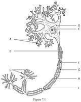
Q376. Using Figure 7.1, identify the neural processes that convey incoming messages toward the cell body.
Background
Topic: Neuron Structure
This question tests your understanding of neuron anatomy, specifically the dendrites, which receive signals and transmit them toward the cell body.
Key Terms:
Dendrites: Branch-like extensions that receive incoming signals.
Cell body (soma): The metabolic center of the neuron.
Step-by-Step Guidance
Look at Figure 7.1 and find the structures that branch out from the cell body.
Identify which label points to these branching processes.
Recall that dendrites are responsible for receiving signals and transmitting them toward the soma.
Try solving on your own before revealing the answer!
Final Answer: Label B
The neural processes that convey incoming messages toward the cell body are indicated by Label B in Figure 7.1. These are the dendrites.
Q288. Using Figure 8.1, identify the tympanic membrane.
Background
Topic: Anatomy of the Ear
This question tests your ability to locate the tympanic membrane (eardrum), which separates the external ear from the middle ear and vibrates in response to sound waves.
Key Terms:
Tympanic membrane: Thin membrane that transmits sound vibrations.
Middle ear: Contains ossicles and is separated from the external ear by the tympanic membrane.
Step-by-Step Guidance
Examine Figure 8.1 and locate the membrane that forms the boundary between the external and middle ear.
Identify which label points to this thin, oval-shaped structure.
Recall the anatomical position of the tympanic membrane relative to the auditory canal and ossicles.
Try solving on your own before revealing the answer!
Final Answer: Label D
The tympanic membrane is indicated by Label D in Figure 8.1. It is the eardrum that vibrates in response to sound waves.
Q377. Using Figure 7.1, identify the metabolic center of the neuron.
Background
Topic: Neuron Structure
This question tests your knowledge of the cell body (soma), which contains the nucleus and most organelles, and is responsible for the neuron's metabolic activities.
Key Terms:
Cell body (soma): Central part of the neuron containing the nucleus.
Nucleus: Contains genetic material and controls cell functions.
Step-by-Step Guidance
Look at Figure 7.1 and find the large, central structure from which dendrites and axon extend.
Identify which label points to this region.
Recall that the metabolic center is the cell body, not the axon or dendrites.
Try solving on your own before revealing the answer!
Final Answer: Label D
The metabolic center of the neuron is indicated by Label D in Figure 7.1. This is the cell body (soma).
Q289. Using Figure 8.1, identify the semicircular canals.
Background
Topic: Anatomy of the Ear
This question tests your ability to locate the semicircular canals, which are part of the inner ear and are involved in balance and equilibrium.
Key Terms:
Semicircular canals: Three looped structures in the inner ear.
Inner ear: Contains structures for hearing and balance.
Step-by-Step Guidance
Examine Figure 8.1 and locate the looped structures near the cochlea.
Identify which label points to these loops.
Recall that the semicircular canals are positioned superiorly and posteriorly in the inner ear.
Try solving on your own before revealing the answer!
Final Answer: Label C
The semicircular canals are indicated by Label C in Figure 8.1. These structures are responsible for detecting rotational movements of the head.
Q378. Using Figure 7.1, identify the axon terminals.
Background
Topic: Neuron Structure
This question tests your understanding of the axon terminals, which are the endpoints of the axon where neurotransmitters are released.
Key Terms:
Axon terminals: Endings of the axon that release neurotransmitters.
Synapse: Junction between neurons where communication occurs.
Step-by-Step Guidance
Look at Figure 7.1 and find the structures at the end of the axon, opposite the cell body.
Identify which label points to these terminal branches.
Recall that axon terminals are responsible for transmitting signals to other neurons or effectors.
Try solving on your own before revealing the answer!
Final Answer: Label I
The axon terminals are indicated by Label I in Figure 7.1. These are the sites where neurotransmitters are released.
