 Indietro
Structure, Growth, and Function of Bones in Anatomy & Physiology
Guida di studio - Note intelligenti
 Appunti personalizzati basati sui tuoi materiali, ampliati con definizioni chiave, esempi e contesto.
Module 6.1: The Skeletal System and Its Divisions
Axial and Appendicular Divisions
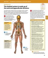
The skeletal system is divided into the axial and appendicular divisions, each serving distinct structural and functional roles.
Axial Skeleton: Composed of the skull, vertebral column, and thoracic cage. Provides support and protection for the brain, spinal cord, and organs in the thorax.
Appendicular Skeleton: Includes the limbs and girdles (pectoral and pelvic). Facilitates movement and interaction with the environment.
Functions of the Skeletal System: Support, protection, movement, storage of minerals, and blood cell production.
Module 6.2: Bone Classification and Markings
Bone Shapes and Structures
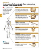
Bones are classified by their shape and structure, which relate to their function and location.
Long Bones: Longer than they are wide; found in limbs (e.g., femur).
Short Bones: Nearly equal in length and width; found in wrists and ankles.
Flat Bones: Thin and broad; found in skull, ribs, and scapulae.
Irregular Bones: Complex shapes; found in vertebrae and pelvis.
Sesamoid Bones: Small, round, and embedded in tendons (e.g., patella).
Bone Type | Description | Example |
|---|---|---|
Long | Longer than wide | Femur |
Short | Nearly equal dimensions | Carpals |
Flat | Thin, broad | Skull |
Irregular | Complex shape | Vertebrae |
Sesamoid | Small, round, in tendons | Patella |
Module 6.3: Bone Markings and Anatomy
Bone Markings
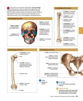
Bone markings are features on bones that serve as sites for muscle attachment, passage of nerves and blood vessels, and articulation with other bones.
Foramen: Opening for blood vessels and nerves.
Process: Projection for muscle attachment.
Condyle: Rounded articular surface.
Fossa: Shallow depression.
Module 6.4: Structure of Long Bones
Long Bone Anatomy
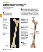
Long bones transmit forces along their shaft and have a rich blood supply.
Diaphysis: Shaft of the bone, composed of compact bone.
Epiphysis: Ends of the bone, composed of spongy bone.
Metaphysis: Region between diaphysis and epiphysis.
Medullary Cavity: Central cavity containing bone marrow.
Periosteum: Outer fibrous layer covering bone.
Endosteum: Inner lining of the medullary cavity.
Module 6.5: Bone Matrix and Cells
Bone Matrix and Cellular Components
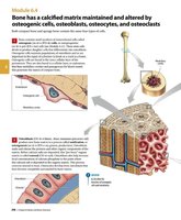
Bone has a calcified matrix maintained and altered by osteogenic cells, osteoblasts, osteocytes, and osteoclasts.
Osteogenic Cells: Stem cells that differentiate into osteoblasts.
Osteoblasts: Cells responsible for bone formation.
Osteocytes: Mature bone cells that maintain bone tissue.
Osteoclasts: Cells that break down bone matrix.
Bone Matrix: Composed of collagen fibers and hydroxyapatite crystals, providing strength and rigidity.
Module 6.6: Compact and Spongy Bone Structure
Osteons and Trabeculae
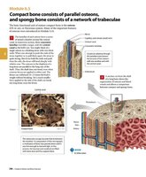
Compact bone consists of parallel osteons, while spongy bone consists of a network of trabeculae.
Osteon (Haversian System): Structural unit of compact bone, containing concentric lamellae around a central canal.
Trabeculae: Lattice-like network in spongy bone, providing structural support and space for marrow.
Canaliculi: Small channels connecting osteocytes for nutrient and waste exchange.
Module 6.7: Bone Growth and Development
Endochondral Ossification
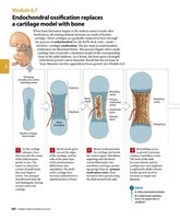
Endochondral ossification replaces a cartilage model with bone, a process essential for the formation of most bones.
Cartilage Model: Initial structure in embryonic development.
Primary Ossification Center: Region where bone formation begins.
Secondary Ossification Center: Develops in epiphyses after birth.
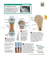
Epiphyseal Plate: Growth plate responsible for lengthening bones.
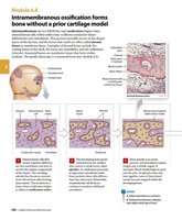
Intramembranous Ossification
Intramembranous ossification forms bone without a prior cartilage model, primarily in flat bones.
Mesenchymal Cells: Differentiate directly into osteoblasts.
Formation of Bone Matrix: Osteoblasts secrete matrix, which calcifies.
Development of Trabeculae: Network forms within the ossifying tissue.
Module 6.8: Appositional Bone Growth
Growth Involving Periosteum and Endosteum
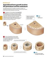
Appositional bone growth increases bone diameter and involves the periosteum and endosteum.
Periosteum: Outer layer where osteoblasts add new bone tissue.
Endosteum: Inner layer where osteoclasts remove bone tissue.
Bone Remodeling: Continuous process of bone formation and resorption.
Module 6.9: Bone Growth at the Epiphyseal Plate
Epiphyseal Plate and Longitudinal Growth
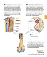
The epiphyseal plate is responsible for the longitudinal growth of bones during childhood and adolescence.
Zones of the Epiphyseal Plate: Resting, proliferating, hypertrophic, and ossification zones.
Closure of Plate: Occurs when bone replaces cartilage, ending growth.
Module 6.10: Bone Development in the Fetus
Fetal Bone Formation
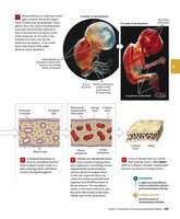
Bone development in the fetus involves both endochondral and intramembranous ossification.
Mesenchymal Cells: Differentiate into chondrocytes or osteoblasts.
Formation of Cartilage Model: Precedes endochondral ossification.
Development of Spongy Bone: Occurs in both processes.
Module 6.11: Bone Remodeling and Repair
Bone Remodeling
Bone remodeling is a lifelong process involving the resorption and formation of bone tissue.
Osteoclasts: Break down old bone.
Osteoblasts: Form new bone.
Importance: Maintains bone strength and mineral homeostasis.
Bone Repair
Bone repair occurs after fractures and involves several stages:
Hematoma Formation: Blood clot forms at the fracture site.
Callus Formation: Cartilage and bone form a temporary bridge.
Bone Remodeling: Restores original bone structure.
Additional info:
Bone tissue is highly vascularized, allowing for efficient nutrient delivery and waste removal.
Calcium and phosphate are the primary minerals stored in bone.
Bone markings are essential for identifying muscle attachment sites and joint articulations.
