 Indietro
Study Guide: Anatomy and Physiology of the Respiratory System
Guida di studio - Note intelligenti
 Appunti personalizzati basati sui tuoi materiali, ampliati con definizioni chiave, esempi e contesto.
The Respiratory System
Overview and Functions
The respiratory system is an organ system responsible for the intake and expulsion of air, facilitating gas exchange, communication, olfaction, acid-base balance, blood pressure regulation, blood and lymph flow, platelet production, blood filtration, and expulsion of abdominal contents. It is essential for maintaining homeostasis and supporting cellular metabolism.
Gas exchange: Oxygen (O2) and carbon dioxide (CO2) are exchanged between blood and air.
Communication: Enables speech and vocalization.
Olfaction: Provides the sense of smell.
Acid-base balance: Regulates pH by eliminating CO2.
Blood pressure regulation: Assists in synthesis of angiotensin II.
Blood and lymph flow: Breathing creates pressure gradients aiding circulation.
Platelet production: Lungs produce more than half of platelets via megakaryocytes.
Blood filtration: Lungs filter small clots.
Expulsion of abdominal contents: Breath-holding assists in urination, defecation, and childbirth.
Organization of Respiratory Organs
The respiratory system is organized into principal organs and functional zones:
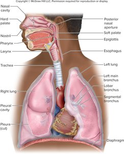
Principal organs: Nose, pharynx, larynx, trachea, bronchi, lungs.
Conducting zone: Passages for airflow only (nostrils to major bronchioles).
Respiratory zone: Regions for gas exchange (alveoli and nearby structures).
Upper respiratory tract: Nose through larynx.
Lower respiratory tract: Trachea through lungs.
The Nose
Structure and Function
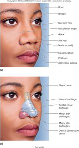
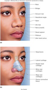
The nose warms, cleanses, and humidifies inhaled air, detects odors, and amplifies voice. It is divided by the nasal septum into right and left nasal fossae, and consists of bone and hyaline cartilage.
Nasal septum: Vomer (inferior), perpendicular plate of ethmoid (superior), septal cartilage (anterior).
Roof: Ethmoid and sphenoid bones.
Floor: Hard palate, separates nasal and oral cavities.
Vestibule: Chamber inside nostrils, lined with stratified squamous epithelium and guard hairs (vibrissae).
Nasal conchae: Superior, middle, and inferior folds projecting from lateral walls; meatuses are narrow passages beneath each concha.
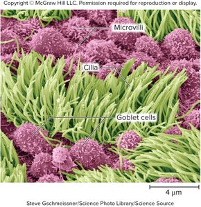
Nasal mucosa: Ciliated pseudostratified columnar epithelium with goblet cells producing mucus.
Olfactory epithelium: Located at roof of nasal fossa, contains immobile cilia for odorant binding.
Lamina propria: Contains large blood vessels for warming air and erectile tissue for alternating airflow.
Anatomy of the Upper Respiratory Tract
Regions and Structures
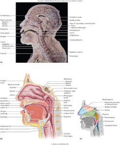
The upper respiratory tract includes the nasal cavity, pharynx, and larynx. It is responsible for filtering, warming, and humidifying air, as well as facilitating speech and swallowing.
Pharynx: Muscular funnel divided into nasopharynx, oropharynx, and laryngopharynx.
Nasopharynx: Receives auditory tubes, contains pharyngeal tonsil, lined by pseudostratified columnar epithelium.
Oropharynx and laryngopharynx: Pass air, food, and drink; lined by stratified squamous epithelium.
Muscles: Assist in swallowing and speech.
The Larynx
Structure and Function
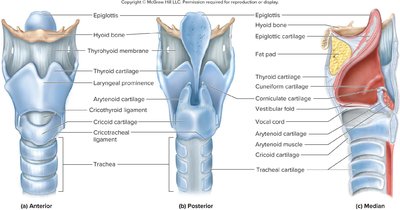
The larynx is a cartilaginous chamber that keeps food and drink out of the airway and produces sound. It is commonly called the "voice box."
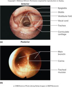
Epiglottis: Flap guarding the superior opening; closes airway during swallowing.
Vestibular folds: Close larynx during swallowing.
Nine cartilages: Epiglottic, thyroid (largest, forms Adam's apple), cricoid (connects larynx to trachea), and others.
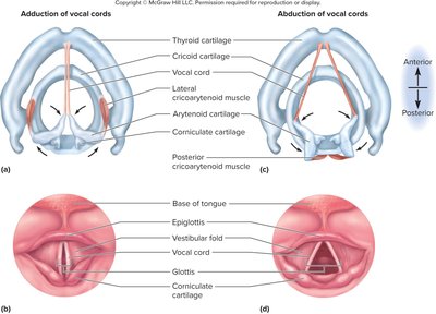
Intrinsic and extrinsic muscles: Control elevation and movement of vocal cords.
Vocal cords: Produce sound when air passes between them; glottis is the opening between cords.
Adult male vocal cords: Longer and thicker, vibrate more slowly, produce lower-pitched sound.
The Trachea
Structure and Histology
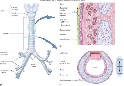
The trachea is a rigid tube connecting the larynx to the bronchi, supported by C-shaped rings of hyaline cartilage. It is lined by ciliated pseudostratified columnar epithelium.
Mucociliary escalator: Mucus traps particles; cilia move mucus to pharynx for swallowing.
Connective tissue: Contains lymphoid nodules, mucous and serous glands, and cartilage.
The Lungs and Bronchial Tree
Gross Anatomy
The lungs are conical organs with distinct landmarks and lobes. The right lung is shorter and has three lobes; the left lung is taller, has two lobes, and a cardiac impression.
Base: Rests on diaphragm.
Apex: Projects above clavicle.
Costal surface: Pressed against ribcage.
Mediastinal surface: Faces heart.
Hilum: Entry for bronchi, blood vessels, lymphatics, nerves.
Bronchial Tree
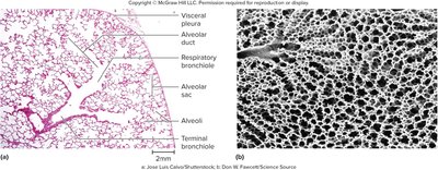
The bronchial tree is a branching system of air tubes extending from the main bronchi to terminal bronchioles.
Main bronchi: Right is wider and more vertical; foreign objects lodge here more often.
Lobar bronchi: Serve each lung lobe.
Segmental bronchi: 10 in right lung, 8 in left lung; ventilate bronchopulmonary segments.
Bronchioles: Lack cartilage, 1 mm or less in diameter; terminal bronchioles are final branches of conducting zone.
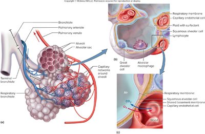
Respiratory bronchioles: Have alveoli budding from walls; begin respiratory zone.
Alveolar ducts and sacs: End in clusters of alveoli around a central atrium.
Alveoli and Respiratory Membrane
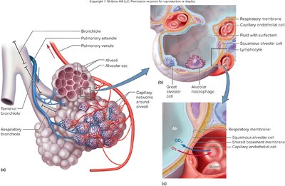
Alveoli are microscopic air pouches (0.2–0.5 mm diameter) where gas exchange occurs. Each alveolus is surrounded by capillaries supplied by the pulmonary artery.
Respiratory membrane: Thin barrier (0.5 µm) between alveolar air and blood, consisting of squamous alveolar cells, endothelial cells of capillary, and shared basement membrane.
The Pleurae
Structure and Function
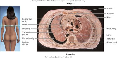
The pleura is a serous membrane lining the thoracic wall and forming the surface of the lung. The pleural cavity is the potential space between pleurae, containing pleural fluid.
Visceral pleura: Covers lung surface.
Parietal pleura: Adheres to mediastinum, rib cage, diaphragm.
Pleural cavity: Contains slippery fluid for reducing friction, creating pressure gradient, and compartmentalization.
Pulmonary Ventilation
Respiratory Cycle and Muscles
Breathing is a repetitive cycle of inspiration and expiration. The flow of air depends on pressure differences created by respiratory muscles.
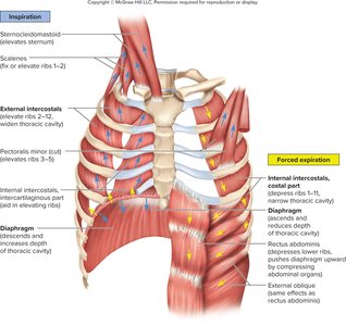
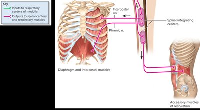
Diaphragm: Prime mover; contraction enlarges thoracic cavity, relaxation compresses lungs.
Intercostal muscles: Assist diaphragm, contribute to thoracic cage movement.
Accessory muscles: Used in forced respiration (e.g., sternocleidomastoid, scalenes, pectoralis major/minor).
Normal quiet expiration: Passive, due to elasticity of lungs and thoracic cage.
Forced expiration: Uses abdominal and other muscles to increase thoracic pressure.
Neural Control of Breathing
Brainstem Respiratory Centers
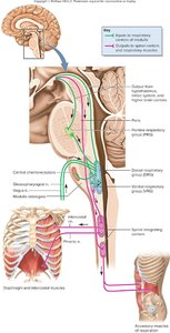
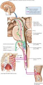
Breathing is controlled by both conscious and unconscious mechanisms. Automatic breathing is regulated by respiratory centers in the medulla oblongata and pons.
Central chemoreceptors: Respond to pH changes in cerebrospinal fluid, reflecting CO2 levels.
Peripheral chemoreceptors: Located in carotid and aortic bodies; respond to O2, CO2, and pH in blood.
Voluntary control: Originates in motor cortex, bypasses brainstem, overridden when CO2 levels rise.
Pressure, Resistance, and Airflow
Principles and Laws
Airflow in the respiratory system is governed by principles of flow, pressure, and resistance. Boyle’s law states that at constant temperature, the pressure of a gas is inversely proportional to its volume:
Boyle’s law:
Atmospheric pressure: 760 mm Hg at sea level (1 atm).
Intrapulmonary pressure: Changes with lung volume.
Intrapleural pressure: Slightly negative, keeps lungs expanded.
Inspiration: Lung volume increases, pressure drops, air flows in.
Expiration: Lung volume decreases, pressure rises, air flows out.
Resistance to Airflow
Bronchiole diameter: Bronchodilation increases airflow; bronchoconstriction decreases airflow.
Pulmonary compliance: Ease of lung expansion; reduced by diseases and surface tension.
Surfactant: Reduces surface tension in alveoli; deficiency causes infant respiratory distress syndrome (IRDS).
Composition of Air and Gas Exchange
Partial Pressures and Laws
Air is a mixture of gases; each gas exerts a partial pressure. Dalton’s law states that total atmospheric pressure is the sum of individual gas pressures. Henry’s law describes gas solubility in liquids:
Dalton’s law:
Henry’s law: Amount of gas dissolved in liquid is proportional to its partial pressure and solubility.
Alveolar Gas Exchange
Pressure gradients: Drive diffusion of O2 and CO2 across respiratory membrane.
Solubility: CO2 is 20 times more soluble than O2.
Membrane surface area and thickness: Affect efficiency; diseases can reduce surface area or increase thickness.
Ventilation–perfusion coupling: Matches airflow and blood flow for optimal gas exchange.
Gas Transport
Oxygen Transport
Hemoglobin carries oxygen in the blood. Each molecule can bind up to four O2 molecules. The oxyhemoglobin dissociation curve illustrates the relationship between hemoglobin saturation and ambient PO2.
Oxyhemoglobin dissociation curve: Not linear; shows rapid loading at higher PO2.
Pulse oximeter: Measures oxygen saturation (SpO2); normal is 95–100%.
Carbon monoxide poisoning: CO binds hemoglobin 210 times more tightly than O2, causing hypoxia.
Respiratory Pathologies
Chronic Obstructive Pulmonary Diseases (COPD)
Chronic bronchitis: Inflammation, excess mucus, impaired cilia, chronic cough, hypoxemia, cyanosis.
Emphysema: Alveolar walls break down, reduced gas exchange, lungs less elastic, air trapped, barrel chest.
Other effects: Reduced vital capacity, hypoxemia, hypercapnia, respiratory acidosis, polycythemia, cor pulmonale.
Lung Cancer
Smoking: Major cause; 90% originate in mucus membranes of large bronchi.
Tumor: Invades bronchial wall, compresses airway, rapid metastasis, poor prognosis.
Additional info: Academic context was added to clarify the structure, function, and clinical relevance of each anatomical region and physiological process. Tables and equations were included where appropriate to reinforce key concepts. Images were selected strictly for their direct relevance to the adjacent content, supporting anatomical and physiological explanations.
