 Indietro
Study Guide: Endocrine Organs, Blood, and Blood Vessels
Guida di studio - Note intelligenti
 Appunti personalizzati basati sui tuoi materiali, ampliati con definizioni chiave, esempi e contesto.
Endocrine Organs
Overview of the Endocrine System
The endocrine system is composed of glands that secrete hormones directly into the bloodstream, regulating various physiological processes such as metabolism, growth, and reproduction. Endocrine glands include the hypothalamus, pituitary gland, thyroid gland, parathyroid glands, adrenal glands, pancreas, pineal gland, and organs with secondary endocrine functions (heart, thymus, digestive tract, adipose tissue, kidneys, gonads).
Hormones: Chemical messengers that influence target organs.
Primary endocrine organs: Hypothalamus, pituitary, thyroid, parathyroid, adrenal, pancreas, pineal.
Secondary endocrine functions: Heart, thymus, digestive tract, adipose tissue, kidneys, gonads.
Functions: Regulation of blood pressure, immunity, digestion, metabolism, and reproduction.
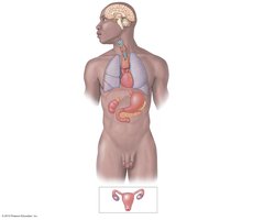
Hypothalamus and Pituitary Gland
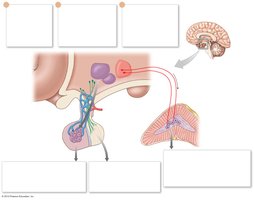
The hypothalamus is a key regulatory center in the brain, controlling the pituitary gland through neural and hormonal signals. The pituitary gland is divided into anterior and posterior lobes, each releasing different hormones.
Hypothalamus: Synthesizes antidiuretic hormone (ADH) and oxytocin; releases regulatory hormones targeting the anterior pituitary.
Anterior pituitary: Releases hormones affecting thyroid, adrenal cortex, and reproductive organs.
Posterior pituitary: Releases ADH and oxytocin into circulation.
Adrenal medulla: Direct neural control by hypothalamus; secretes epinephrine and norepinephrine.
Thyroid and Parathyroid Glands
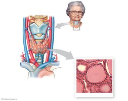
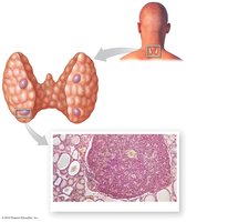
The thyroid gland is located in the neck and produces hormones that regulate metabolism. The parathyroid glands are embedded in the thyroid and regulate calcium levels.
Thyroid gland: Composed of follicles lined by simple cuboidal epithelium; produces thyroxine (T4) and triiodothyronine (T3).
Parathyroid glands: Small glands on the posterior thyroid; secrete parathyroid hormone (PTH) to increase blood calcium.
Histology: Thyroid follicles contain colloid with thyroglobulin; C cells produce calcitonin.
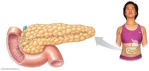
Pancreas and Pancreatic Islets
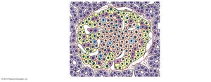
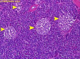
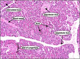
The pancreas has both endocrine and exocrine functions. The endocrine portion consists of pancreatic islets (islets of Langerhans), which regulate blood glucose.
Alpha cells: Produce glucagon, raising blood glucose.
Beta cells: Produce insulin, lowering blood glucose.
Pancreatic acini: Exocrine cells producing digestive enzymes.
Pineal Gland
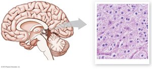
The pineal gland is located in the brain and secretes melatonin, which regulates circadian rhythms.
Melatonin: Hormone involved in sleep-wake cycles.
Adrenal Glands
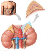
The adrenal glands are located atop the kidneys and consist of an outer cortex and inner medulla.
Adrenal cortex: Produces mineralocorticoids (aldosterone), glucocorticoids (cortisol), and androgens.
Adrenal medulla: Produces epinephrine and norepinephrine.
Hormonal effects: Regulation of sodium, glucose metabolism, inflammation, and sympathetic responses.
Blood
Components of Blood
Blood is a connective tissue composed of plasma and formed elements.
Plasma: Liquid matrix containing water, proteins (albumins, globulins, fibrinogen), electrolytes, nutrients, and wastes.
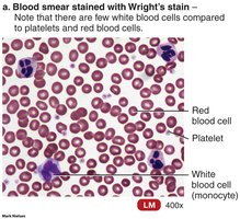
Formed elements: Red blood cells (RBCs), white blood cells (WBCs), and platelets.
Red Blood Cells (Erythrocytes)
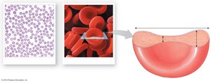
RBCs are biconcave cells responsible for oxygen transport.
Structure: Biconcave shape increases surface area for gas exchange.
Function: Carry oxygen via hemoglobin; remove carbon dioxide.
White Blood Cells (Leukocytes)
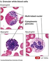
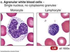
WBCs are involved in immune defense and are classified as granular or agranular.
Granular leukocytes: Neutrophils, eosinophils, basophils.
Agranular leukocytes: Monocytes, lymphocytes.
Functions: Phagocytosis, antibody production, inflammation, defense against pathogens.
Blood Types and Antigens
Blood types are determined by surface antigens on RBCs and corresponding antibodies in plasma.
Type A: Surface antigen A, anti-B antibodies.
Type B: Surface antigen B, anti-A antibodies.
Type AB: Both antigens, no antibodies.
Type O: No antigens, both antibodies.
Agglutination and Hemolysis
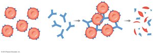
Agglutination occurs when antibodies react with incompatible antigens, causing clumping and destruction of RBCs.
Agglutination: Clumping of RBCs due to antibody-antigen reaction.
Hemolysis: Destruction of RBCs following agglutination.
Blood Vessels
Structure of Blood Vessels
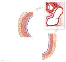
Blood vessels are classified as arteries, veins, and capillaries, each with distinct structural layers.
Tunica intima: Inner endothelial layer.
Tunica media: Middle layer of smooth muscle and elastic fibers.
Tunica externa: Outer connective tissue layer.
Types of Arteries and Veins
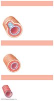
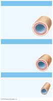
Arteries are classified as elastic, muscular, and arterioles; veins as large, medium-sized, and venules.
Elastic arteries: Large vessels with abundant elastic fibers.
Muscular arteries: Medium-sized, more smooth muscle.
Arterioles: Smallest arteries, regulate blood flow to capillaries.
Veins: Contain valves to prevent backflow; thinner walls than arteries.
Venous Valves and Blood Flow
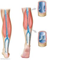
Venous valves ensure unidirectional blood flow toward the heart, especially in limbs.
Valves: Prevent backflow of blood.
Muscle contraction: Helps propel blood through veins.
Capillary Beds and Blood Flow Regulation
Capillary beds are networks of capillaries that regulate blood flow via precapillary sphincters and arteriovenous anastomoses.
Precapillary sphincters: Control blood flow into capillaries.
Arteriovenous anastomosis: Direct connection between arterioles and venules.
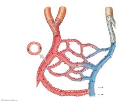
Circuits of the Cardiovascular System
The cardiovascular system consists of the pulmonary and systemic circuits.
Pulmonary circuit: Carries blood from the heart to the lungs and back.
Systemic circuit: Carries blood from the heart to the rest of the body.
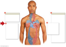
Major Arteries and Veins
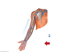
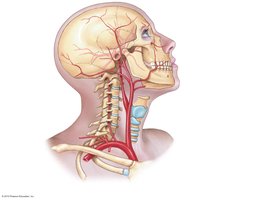
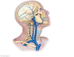
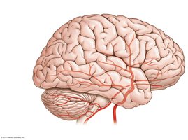
The body contains numerous arteries and veins, each serving specific regions.
Arteries: Carry blood away from the heart; include aorta, carotid, subclavian, femoral, etc.
Veins: Return blood to the heart; include jugular, subclavian, femoral, saphenous, etc.
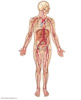
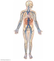
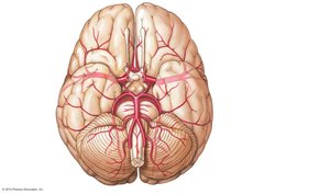
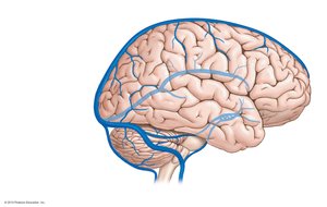
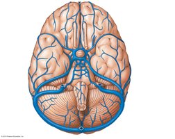
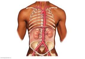
Arteries and Veins of the Lower Limb
The lower limb is supplied by the femoral, popliteal, tibial, and fibular arteries and drained by corresponding veins.
Arteries: Dorsalis pedis, plantar arch, femoral, popliteal, tibial, fibular.
Veins: Great saphenous, small saphenous, femoral, popliteal, tibial, fibular.
Summary Table: Adrenal Hormones
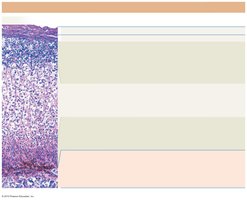
The following table summarizes the regions of the adrenal gland, their hormones, targets, effects, and regulatory control:
Region/Zone | Hormones | Primary Targets | Hormonal Effects | Regulatory Control |
|---|---|---|---|---|
Outer zone of cortex | Mineralocorticoids (aldosterone) | Kidneys | Increased Na+ and water reabsorption; accelerates K+ loss | Stimulated by renin-angiotensin system; inhibited by opposing hormones |
Central zone of cortex | Glucocorticoids (cortisol) | Most cells | Increase glucose/glycogen formation; reduce inflammation | Stimulated by ACTH |
Narrow zone bordering medulla | Androgens | Skin, bones, other tissues | Minimal effects in adults; stimulate pubic hair development pre-puberty | Stimulated by ACTH |
Adrenal medulla | Epinephrine, norepinephrine | Most cells | Increase cardiac activity, blood pressure, glycogen breakdown, blood glucose | Stimulated by sympathetic preganglionic fibers |
Key Equations
Osmosis and hormone action:
Blood flow: (where Q = flow, \Delta P = pressure difference, R = resistance)
Additional info: Academic context was added to clarify hormone functions, blood vessel structure, and blood cell classification.
