 Indietro
Study Guide: Joints (Articulations) in Human Anatomy & Physiology
Guida di studio - Note intelligenti
 Appunti personalizzati basati sui tuoi materiali, ampliati con definizioni chiave, esempi e contesto.
Joints (Articulations)
Introduction to Joints
Joints, also known as articulations, are the sites where two or more bones meet. They play a crucial role in providing mobility to the skeleton and holding it together. Understanding the classification and structure of joints is essential for comprehending movement and stability in the human body.
Classification of Joints
Structural Classification
Fibrous Joints: Bones joined by dense fibrous connective tissue; no joint cavity; mostly immovable.
Cartilaginous Joints: Bones united by cartilage; no joint cavity; not highly movable.
Synovial Joints: Bones separated by a fluid-filled joint cavity; all are freely movable.
Functional Classification
Synarthroses: Immovable joints
Amphiarthroses: Slightly movable joints
Diarthroses: Freely movable joints
Structural classifications are more clear-cut and are commonly used in anatomical study.
Fibrous Joints
General Features
Fibrous joints are characterized by bones joined by dense fibrous connective tissue, lacking a joint cavity. Most are immovable, but the degree of movement depends on the length of connective tissue fibers.
Types of Fibrous Joints
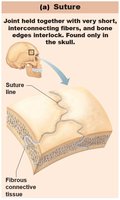
Sutures: Very short, interconnecting fibers; bone edges interlock; found only in the skull.
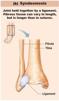
Syndesmoses: Bones connected by ligaments; fiber length varies, so movement varies.
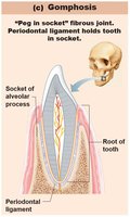
Gomphoses: Peg-in-socket joints; only examples are teeth in alveolar sockets, held by the periodontal ligament.
Syndesmoses
Connected by ligaments (bands of fibrous tissue).
Short fibers offer little to no movement (e.g., inferior tibiofibular joint).
Longer fibers allow more movement (e.g., interosseous membrane between radius and ulna).
Gomphoses
Peg-in-socket fibrous joint.
Periodontal ligament holds the tooth in its socket.
Cartilaginous Joints
General Features
Cartilaginous joints unite bones with cartilage and lack a joint cavity. They are not highly movable and are classified into two types: synchondroses and symphyses.
Types of Cartilaginous Joints
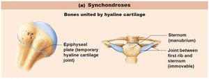
Synchondroses: Bar or plate of hyaline cartilage unites bones; almost all are immovable.
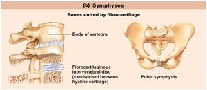
Symphyses: Fibrocartilage unites bones; hyaline cartilage also present as articular cartilage; strong, slightly movable joints.
Synchondroses
Examples: Temporary epiphyseal plate joints (become synostoses after closure), cartilage of 1st rib with manubrium of sternum.
Symphyses
Examples: Intervertebral joints, pubic symphysis.
Fibrocartilage provides strength and flexibility.
Synovial Joints
General Features
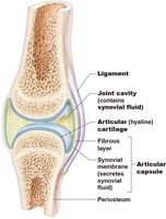
Synovial joints are characterized by bones separated by a fluid-filled joint cavity. All synovial joints are diarthrotic (freely movable) and include almost all limb joints. They have six general features and are associated with bursae and tendon sheaths.
Articular cartilage: Hyaline cartilage covering bone ends; prevents crushing.
Joint (synovial) cavity: Small, fluid-filled space unique to synovial joints.
Articular (joint) capsule: Two layers: external fibrous layer (dense irregular connective tissue) and inner synovial membrane (loose connective tissue).
Synovial fluid: Viscous, slippery filtrate of plasma and hyaluronic acid; lubricates and nourishes articular cartilage; contains phagocytic cells.
Bursae and Tendon Sheaths
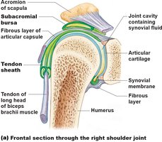
Bursae: Bags of synovial fluid that reduce friction where ligaments, muscles, skin, tendons, or bones rub together.
Tendon sheaths: Elongated bursae wrapped around tendons subjected to friction.
Movements Allowed by Synovial Joints
Types of Movements
Synovial joints allow a variety of movements, classified as gliding, angular, and rotational. Special movements also occur at certain joints.
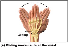
Gliding: One flat bone surface glides or slips over another.
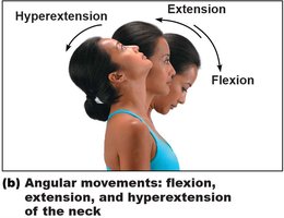
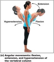
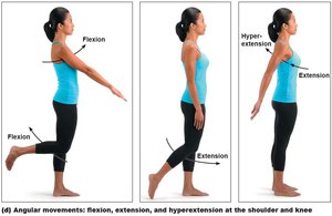
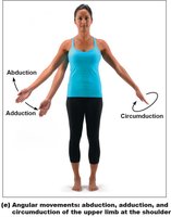
Angular: Increase or decrease the angle between two bones (flexion, extension, hyperextension, abduction, adduction, circumduction).
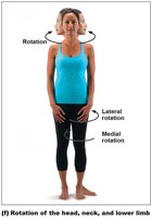
Rotation: Turning of bone around its own long axis (medial and lateral rotation).
Special movements: Supination, pronation, dorsiflexion, plantar flexion, inversion, eversion, protraction, retraction.
Gliding Movements
Examples: Intercarpal joints, intertarsal joints, between articular processes of vertebrae.
Angular Movements
Flexion: Decreases the angle of the joint.
Extension: Increases the angle of the joint.
Hyperextension: Movement beyond the anatomical position.
Abduction, Adduction, Circumduction
Abduction: Movement away from the midline.
Adduction: Movement toward the midline.
Circumduction: Combination of flexion, abduction, extension, and adduction; limb describes a cone in space.
Rotation
Medial rotation: Toward the midline.
Lateral rotation: Away from the midline.
Examples: Rotation between C1 and C2 vertebrae, rotation of humerus and femur.
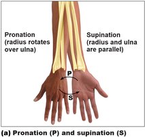
Special Movements
Supination: Palms face anteriorly; radius and ulna are parallel.
Pronation: Palms face posteriorly; radius rotates over ulna.
Dorsiflexion: Bending foot toward shin.
Plantar flexion: Pointing toes.
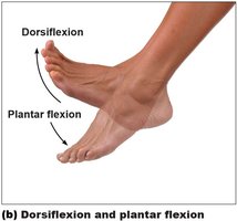
Inversion: Sole of foot faces medially.
Eversion: Sole of foot faces laterally.
Protraction: Mandible juts out.
Retraction: Mandible is pulled toward neck.
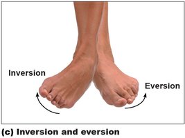
Types of Synovial Joints
Classification by Shape and Movement
Synovial joints are classified into six types based on the shape of their articular surfaces and the movements they allow:
Plane
Hinge
Pivot
Condylar
Saddle
Ball-and-socket
Each type allows specific movements and is found in characteristic locations throughout the body.
Summary Table: Joint Classification
Structural Type | Material/Features | Movement | Examples |
|---|---|---|---|
Fibrous | Dense fibrous connective tissue | Immovable to slightly movable | Sutures (skull), syndesmoses (tibia-fibula), gomphoses (teeth) |
Cartilaginous | Cartilage (hyaline or fibrocartilage) | Immovable to slightly movable | Epiphyseal plate, pubic symphysis, intervertebral discs |
Synovial | Joint cavity with synovial fluid | Freely movable | Shoulder, knee, wrist, hip |
Key Terms: articulation, synarthrosis, amphiarthrosis, diarthrosis, synovial fluid, bursae, tendon sheath, flexion, extension, abduction, adduction, circumduction, rotation, supination, pronation, dorsiflexion, plantar flexion, inversion, eversion, protraction, retraction.
Example: The knee joint is a synovial hinge joint, allowing flexion and extension, stabilized by ligaments and cushioned by synovial fluid.
Additional info: Synovial joints are the most common and most complex type of joint in the human body, enabling a wide range of movements essential for daily activities and athletic performance.
