 Indietro
Study Guide: Lower Appendicular Skeleton – Pelvic Girdle, Lower Limb Bones, Joints, and Muscles
Guida di studio - Note intelligenti
 Appunti personalizzati basati sui tuoi materiali, ampliati con definizioni chiave, esempi e contesto.
Lower Appendicular Skeleton
Overview
The lower appendicular skeleton consists of the pelvic girdle and the lower limb. It plays a crucial role in supporting body weight, locomotion, and protecting internal organs. The bones, joints, and muscles of this region are more robust than those of the upper limb due to their functional demands.
Pelvic Girdle: Supports and protects lower viscera and, in females, the developing fetus.
Lower Limb: Includes the thigh (femur), kneecap (patella), leg (tibia and fibula), ankle (tarsals), and foot (metatarsals and phalanges).
The Pelvic Girdle
Anatomy and Structure
The pelvic girdle is formed by two ossa coxae bones, which unite at the symphysis pubis and sacrum to create a girdle. Each os coxae consists of three bones that fuse at the acetabulum: the ilium, ischium, and pubis.
Os Coxae: Also known as the innominate bone.
Acetabulum: The socket for the femoral head, formed by the union of the three bones.
Pelvic Outlet: The inferior opening of the pelvis, important in childbirth.
Ischial Spine: A bony projection important for pelvic measurements.
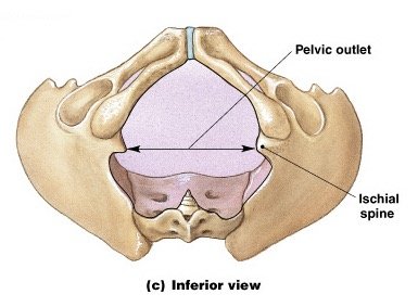
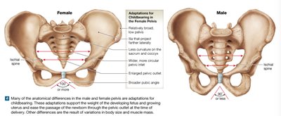
Male vs. Female Pelvis
There are significant anatomical differences between the male and female pelvis, primarily related to childbirth adaptations in females.
Male Pelvis: Heavier, more prominent markings, narrower pelvic inlet, more curved sacrum and coccyx.
Female Pelvis: Enlarged pelvic outlet, less curvature of sacrum and coccyx, wider and more circular pelvic inlet, broader and lower pelvis, widely fanning ilia, pubic angle greater than 100°.
Bones of the Lower Limb
Femur
The femur is the longest and strongest bone in the body, forming the thigh. It articulates proximally with the pelvis and distally with the tibia and patella.
Head: Large, round, with a shallow pit for the ligamentum teres.
Body: Curved from greater trochanter to epicondyles.
Condyles: Articulate with tibia.
Patellar Surface: Articulates with the patella.
Linea Aspera: Posterior ridge for muscle attachment.
Patella
The patella is a sesamoid bone embedded in the quadriceps tendon, protecting the knee joint and improving leverage for knee extension.
Tibia and Fibula
Tibia: The main weight-bearing bone of the leg, with articular condyles separated by the intercondylar eminence and a tibial tuberosity for patellar ligament attachment.
Fibula: Slender, important for muscle attachments, minimal weight support, distal end forms the lateral malleolus.
Joints of the Lower Limb
Hip Joint
The hip joint is a ball-and-socket synovial joint formed by the acetabulum of the pelvis and the head of the femur. It allows for a wide range of motion, including flexion, extension, abduction, adduction, and rotation.
Knee Joint
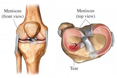
The knee is a complex hinge joint involving the femur, tibia, and patella. It is stabilized by several ligaments and contains menisci for shock absorption and joint congruency.
Menisci: Medial and lateral fibrocartilaginous structures that cushion and stabilize the joint.
Ligaments:
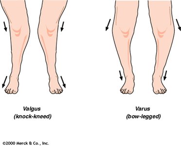
MCL (Medial Collateral Ligament): Prevents valgus (knock-knee) stress.
LCL (Lateral Collateral Ligament): Prevents varus (bow-leg) stress.
ACL (Anterior Cruciate Ligament): Prevents anterior displacement of tibia.
PCL (Posterior Cruciate Ligament): Prevents posterior displacement of tibia.
Oblique/Arcuate Popliteal Ligament, Transverse Ligament, Patellar Ligament: Additional stabilizers.
Muscles of the Lower Limb
Overview
The muscles of the lower limb are larger and more powerful than those of the upper limb, reflecting their role in locomotion and weight-bearing. They are organized into groups based on their actions and anatomical location.
Muscles That Move the Thigh
Posterior (Gluteal) Group
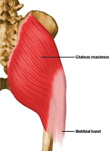
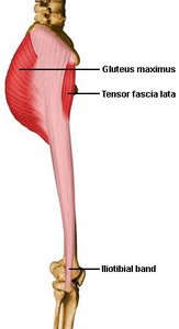
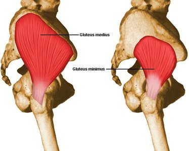
The gluteal region contains the gluteus maximus, medius, and minimus muscles, which are key for hip extension, abduction, and medial rotation.
Gluteus Maximus: Powerful hip extensor, forms the roundness of the buttock, extensive attachments.
Gluteus Medius and Minimus: Lie deeper, abduct and medially rotate the hip.
Deep (Lateral Rotator) Group
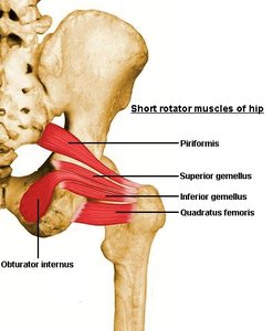
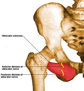
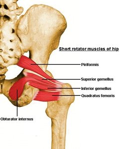
This group consists of six deep muscles that laterally rotate the hip. Most insert on the greater trochanter, and the sciatic nerve passes beneath the piriformis.
Muscles: Piriformis, superior gemellus, inferior gemellus, obturator internus, obturator externus, quadratus femoris.
Medial (Adductor) Group
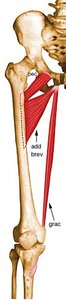
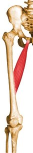
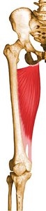
These muscles adduct the hip and may assist in flexion and extension. Most originate from the pubic bone and insert on the femur.
Adductor Brevis, Longus, Magnus: Main adductors, Magnus also originates from the ischium.
Pectineus, Gracilis: Additional adductors.
Muscles That Move the Leg
Extensors
The quadriceps femoris group (vasti and rectus femoris) is the primary extensor of the knee, with a common patellar attachment. The sartorius crosses obliquely, flexing and rotating the hip and knee.
Flexors
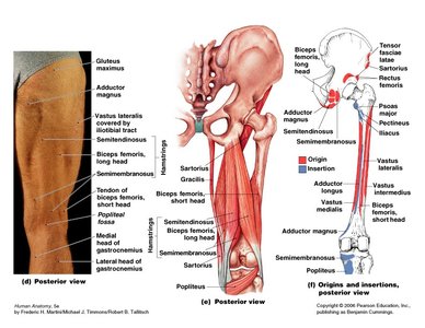
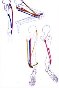
The hamstrings (biceps femoris, semimembranosus, semitendinosus) act to extend and rotate the hip and flex the knee. They are antagonists to the quadriceps and share a common origin on the ischial tuberosity.
Biceps Femoris: Laterally located, only long head arises from ischium.
Semimembranosus and Semitendinosus: Medially located.
Adductor Magnus: A portion acts with the hamstrings.
Summary Table: Key Differences Between Male and Female Pelvis
Feature | Male Pelvis | Female Pelvis |
|---|---|---|
Pelvic Outlet | Narrower | Wider |
Sacrum/Coccyx Curvature | More Curved | Less Curved |
Pelvic Inlet | Narrow, Heart-shaped | Wide, Circular |
Pubic Angle | < 90° | > 100° |
Ilia | Less Fanning | Widely Fanning |
Bone Mass | Heavier | Lighter |
Summary Table: Major Muscles of the Lower Limb
Muscle Group | Primary Muscles | Action |
|---|---|---|
Gluteal | Gluteus maximus, medius, minimus | Hip extension, abduction, medial rotation |
Lateral Rotators | Piriformis, gemelli, obturators, quadratus femoris | Lateral rotation of hip |
Adductors | Adductor brevis, longus, magnus, pectineus, gracilis | Adduction of hip |
Quadriceps | Rectus femoris, vastus lateralis, medialis, intermedius | Knee extension |
Hamstrings | Biceps femoris, semimembranosus, semitendinosus | Hip extension, knee flexion |
Key Equations and Concepts
Joint Stability and Movement
Valgus and Varus:
Valgus: Knock-kneed alignment, prevented by MCL.
Varus: Bow-legged alignment, prevented by LCL.
Meniscus Function: Shock absorption, joint congruency, injury risk (e.g., meniscus tear).
Muscle Actions
Quadriceps: Knee extension
Hamstrings: Knee flexion, hip extension
Adductors: Hip adduction
Gluteal Muscles: Hip extension, abduction, medial rotation
Ligament Functions
MCL: Prevents valgus stress
LCL: Prevents varus stress
ACL: Prevents anterior drawer
PCL: Prevents posterior drawer
Additional info:
Tables referenced (11.11 to 11.16) likely provide detailed muscle origins, insertions, actions, and innervation. These are summarized in the muscle tables above.
Figures referenced (7.1, 7.10a/b, 7.11a/b, 7.12, 7.13, 7.14, 7.15, 8.13-8.16, 11.10, 11.12, 11.16, 11.18, 11.19, 11.21, 11.28) correspond to anatomical diagrams and muscle charts, many of which are visually represented in the included images.
