 Indietro
Study Guide: Muscle Structure and Function (Chapter 12 - Human Physiology)
Guida di studio - Note intelligenti
 Appunti personalizzati basati sui tuoi materiali, ampliati con definizioni chiave, esempi e contesto.
Q1. Identify and describe the parts of the sarcomere.
Background
Topic: Skeletal Muscle Structure
This question tests your understanding of the anatomy of the sarcomere, the fundamental contractile unit of striated muscle. Knowing the parts and their functions is essential for understanding muscle contraction.
Key Terms:
Sarcomere: The basic unit of muscle contraction, defined by the area between two Z lines.
Actin (thin filament): Protein filament involved in muscle contraction.
Myosin (thick filament): Motor protein responsible for force generation.
Z line: Boundary of the sarcomere.
M line: Center of the sarcomere.
A band: Region containing the entire length of the thick filaments.
I band: Region containing only thin filaments.
H zone: Region in the center of the A band with only thick filaments.
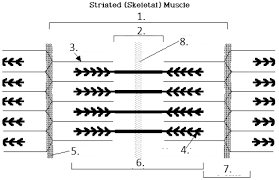
Step-by-Step Guidance
Examine the diagram and locate the boundaries of the sarcomere, which are marked by the Z lines.
Identify the thick filaments (myosin) and thin filaments (actin) within the sarcomere.
Find the A band, which spans the length of the thick filaments, and the I band, which contains only thin filaments.
Locate the H zone, which is the central region of the A band where only thick filaments are present.
Identify the M line, which is the center of the sarcomere and anchors the thick filaments.
Review the diagram and match each labeled part to its anatomical name and function, using the key terms above.
Try solving on your own before revealing the answer!
Final Answer:
1. Sarcomere (area between two Z lines)
2. A band (length of thick filaments)
3. Thin filament (actin)
4. Thick filament (myosin)
5. Z line (boundary of sarcomere)
6. H zone (center of A band, only thick filaments)
7. I band (only thin filaments)
8. M line (center of sarcomere, anchors thick filaments)
Each part plays a specific role in muscle contraction, with the arrangement of actin and myosin filaments allowing for the sliding filament mechanism.
Q2. What is the importance of tropomyosin and troponin?
Background
Topic: Regulation of Muscle Contraction
This question tests your understanding of how muscle contraction is regulated at the molecular level, specifically by the proteins tropomyosin and troponin.
Key Terms:
Tropomyosin: A protein that covers myosin-binding sites on actin, preventing contraction in resting muscle.
Troponin: A protein complex that binds calcium ions and moves tropomyosin away from the binding sites, allowing contraction.
Step-by-Step Guidance
Recall that muscle contraction requires myosin to bind to actin.
Understand that tropomyosin blocks these binding sites when the muscle is relaxed.
Troponin, when it binds calcium, changes shape and moves tropomyosin off the binding sites.
This allows myosin to interact with actin, initiating contraction.
Try solving on your own before revealing the answer!
Final Answer:
Tropomyosin prevents muscle contraction by blocking myosin-binding sites on actin. Troponin, when it binds calcium, shifts tropomyosin away, allowing contraction to occur. Together, they regulate muscle contraction in response to calcium levels.
Q3. Describe the events that occur related to the neuromuscular junction. Begin with an action potential as it reaches the axon terminal and end with the neurotransmitter reaching the muscle.
Background
Topic: Neuromuscular Transmission
This question tests your understanding of how nerve impulses are transmitted to muscle fibers, leading to muscle contraction.
Key Terms:
Neuromuscular junction: The synapse between a motor neuron and a muscle fiber.
Action potential: Electrical signal traveling down the neuron.
Acetylcholine (ACh): The neurotransmitter released at the neuromuscular junction.
Synaptic cleft: The gap between the neuron and muscle fiber.
Step-by-Step Guidance
When an action potential reaches the axon terminal, voltage-gated calcium channels open.
Calcium enters the terminal, triggering vesicles containing acetylcholine to fuse with the membrane.
Acetylcholine is released into the synaptic cleft.
Acetylcholine binds to receptors on the muscle fiber membrane (sarcolemma).
Try solving on your own before revealing the answer!
Final Answer:
After the action potential arrives, calcium influx causes acetylcholine release, which crosses the synaptic cleft and binds to muscle fiber receptors, initiating muscle activation.
Q4. Describe the excitation-contraction coupling mechanism: a) Excitation b) Contraction
Background
Topic: Muscle Physiology
This question tests your understanding of how an electrical signal (excitation) leads to muscle contraction (mechanical response).
Key Terms:
Excitation: The process of generating an action potential in the muscle fiber.
Contraction: The process by which muscle fibers shorten and generate force.
Sarcoplasmic reticulum: Organelle that stores calcium.
T tubules: Invaginations of the sarcolemma that help transmit the action potential.
Step-by-Step Guidance
Excitation begins when the action potential travels along the sarcolemma and down the T tubules.
This triggers the release of calcium from the sarcoplasmic reticulum.
Calcium binds to troponin, causing tropomyosin to move and expose myosin-binding sites on actin.
Myosin heads bind to actin, forming cross-bridges and initiating contraction.
Try solving on your own before revealing the answer!
Final Answer:
Excitation involves action potential propagation and calcium release; contraction occurs when calcium enables cross-bridge formation between actin and myosin, resulting in muscle shortening.
Q5. What are the three sources of energy for muscle contraction?
Background
Topic: Muscle Metabolism
This question tests your knowledge of how muscles obtain energy to contract.
Key Terms:
ATP: Immediate energy source for contraction.
Creatine phosphate: Rapidly regenerates ATP.
Cellular respiration: Produces ATP from glucose and fatty acids.
Step-by-Step Guidance
Recall that ATP is required for myosin head movement and detachment.
Creatine phosphate can quickly donate a phosphate to ADP to regenerate ATP.
Cellular respiration (aerobic and anaerobic) provides sustained ATP production.
Try solving on your own before revealing the answer!
Final Answer:
The three sources are: ATP, creatine phosphate, and cellular respiration (glycolysis and oxidative phosphorylation). These provide energy for muscle contraction at different time scales.
Q6. Describe the three types of muscle fibers.
Background
Topic: Muscle Fiber Types
This question tests your understanding of the structural and functional differences between muscle fiber types.
Key Terms:
Slow-twitch (Type I): Fatigue-resistant, aerobic, for endurance.
Fast-twitch (Type IIa): Intermediate, both aerobic and anaerobic.
Fast-twitch (Type IIb): Fatigue quickly, anaerobic, for power.
Step-by-Step Guidance
Identify the characteristics of slow-twitch fibers (Type I): high endurance, rich in mitochondria, use aerobic metabolism.
Describe fast-twitch fibers (Type IIa): intermediate properties, can use both aerobic and anaerobic metabolism.
Describe fast-twitch fibers (Type IIb): generate high force, fatigue quickly, rely on anaerobic metabolism.
Try solving on your own before revealing the answer!
Final Answer:
Type I (slow-twitch) fibers are endurance-oriented; Type IIa (fast-twitch) are intermediate; Type IIb (fast-twitch) are power-oriented and fatigue quickly. Each type is suited for different physical activities.
Q7. Describe the types of reflexes.
Background
Topic: Reflex Physiology
This question tests your understanding of the different types of reflexes in the human body.
Key Terms:
Monosynaptic reflex: Involves a single synapse (e.g., stretch reflex).
Polysynaptic reflex: Involves multiple synapses (e.g., withdrawal reflex).
Muscle spindle: Detects stretch in muscle.
Golgi tendon organ: Detects tension in tendon.
Step-by-Step Guidance
Recall that monosynaptic reflexes involve direct communication between sensory and motor neurons.
Polysynaptic reflexes involve interneurons and more complex pathways.
Understand the role of muscle spindles and Golgi tendon organs in reflexes.
Try solving on your own before revealing the answer!
Final Answer:
Reflexes can be monosynaptic (simple, like the knee-jerk) or polysynaptic (complex, like withdrawal). Muscle spindles and Golgi tendon organs are key sensory receptors involved.
Q8. Compare and contrast how the three types of muscles are stimulated to contract. Skeletal, Cardiac, Smooth. Stimulus? Can it contract without nervous system stimulation? Yes/No? Voluntary or involuntary control?
Background
Topic: Muscle Types and Control
This question tests your understanding of the differences in how skeletal, cardiac, and smooth muscles are stimulated and controlled.
Key Terms:
Skeletal muscle: Voluntary, requires nervous system stimulation.
Cardiac muscle: Involuntary, can contract without nervous system input (pacemaker cells).
Smooth muscle: Involuntary, can contract without nervous system input (hormonal/local factors).
Step-by-Step Guidance
Compare the stimulus for contraction in each muscle type: skeletal (nervous system), cardiac (pacemaker cells), smooth (variety of stimuli).
Determine whether each muscle type can contract without nervous system stimulation.
Identify whether control is voluntary or involuntary for each muscle type.
Try solving on your own before revealing the answer!
Final Answer:
Skeletal: Stimulated by nerves, cannot contract without nervous system, voluntary control.
Cardiac: Stimulated by pacemaker cells, can contract without nervous system, involuntary control.
Smooth: Stimulated by nerves, hormones, or local factors, can contract without nervous system, involuntary control.
Each muscle type has unique mechanisms for stimulation and control, reflecting their roles in the body.
