 Indietro
Study Guide: Nervous System and Nervous Tissue – Ion Gradients, Membrane Potentials, and Neural Signaling
Guida di studio - Note intelligenti
 Appunti personalizzati basati sui tuoi materiali, ampliati con definizioni chiave, esempi e contesto.
Ch. 11 Introduction to the Nervous System and Nervous Tissue
Ions: Sodium and Potassium
The movement of ions across neuronal membranes is fundamental to the function of the nervous system. Two key ions, sodium (Na+) and potassium (K+), are responsible for generating and maintaining membrane potentials.
Electrochemical Gradient: The combined effect of the electrical gradient (ions move toward areas of opposite charge) and the concentration gradient (ions move from areas of high concentration to low concentration).
Electrical Gradient: Drives ions toward areas of opposite charge.
Concentration Gradient: Drives ions from high to low concentration.
Net Ion Flow: If gradients oppose, the stronger gradient determines the direction of ion movement.
Example: For potassium ions (K+), the electrical gradient pulls them into the cell, while the concentration gradient pushes them out.
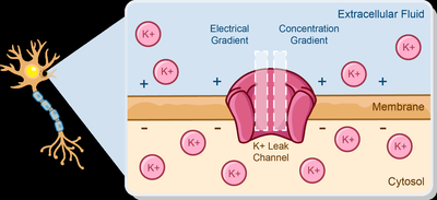
Standard Sodium and Potassium Concentrations
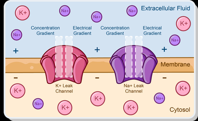
Neurons maintain distinct concentrations of sodium and potassium inside and outside the cell, which is crucial for electrical signaling.
Sodium (Na+): Intracellular concentration is low; extracellular concentration is high.
Potassium (K+): Intracellular concentration is high; extracellular concentration is low.
Example: The concentration and electrical gradients for Na+ and K+ direct their movement across the membrane.
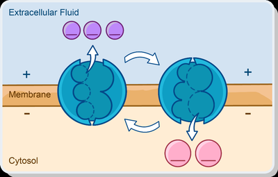
The Sodium-Potassium Pump (Na+/K+ ATPase)
The sodium-potassium pump is an active transport mechanism that maintains the concentration gradients of Na+ and K+ across the membrane.
Function: Uses ATP to move ions against their electrochemical gradients.
Mechanism: Ejects 3 Na+ ions from the cell and transports 2 K+ ions into the cell.
Importance: Maintains resting membrane potential and ionic gradients necessary for action potentials.
Equation:
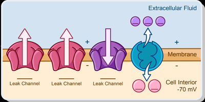
Resting Membrane Potential
The resting membrane potential is the voltage difference across the membrane when the neuron is not actively signaling.
Typical Value: Approximately -70 mV.
Inside of Cell: More negative than the exterior.
Created by: Differences in ionic composition and membrane permeability to ions.
Stabilized by: Na+/K+ ATPase, which maintains concentration gradients.
Change in Membrane Potential
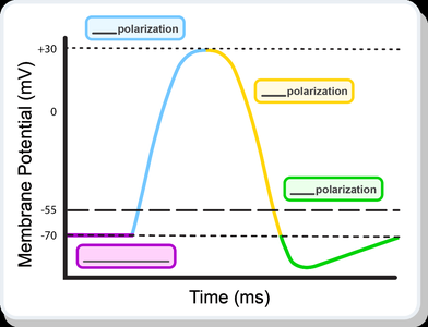
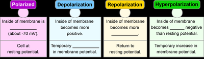
Neurons generate electrical signals by changing their membrane potential. These changes are classified as polarization, depolarization, repolarization, and hyperpolarization.
Polarization: Inside of membrane is negative (resting state).
Depolarization: Membrane potential becomes less negative (more positive).
Repolarization: Return to resting potential after depolarization.
Hyperpolarization: Membrane potential becomes more negative than resting potential.
Example: A neuron’s membrane potential changing from -90 mV to -10 mV is depolarization.
Properties of Graded and Action Potentials
Neurons use two types of electrical signals: graded potentials and action potentials. Each has distinct properties and functions.
Graded Potentials: Variable-strength signals, occur in response to stimuli, mainly in dendrites and cell body.
Action Potentials: Brief, all-or-none depolarization events propagated along the axon.
Threshold: Action potentials initiate at a threshold of approximately -55 mV.
Comparison Table:
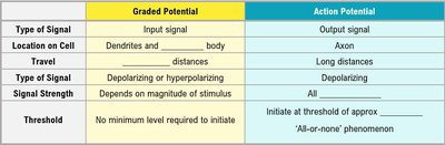
Graded Potential | Action Potential |
|---|---|
Input signal | Output signal |
Dendrites and cell body | Axon |
Short distances | Long distances |
Depolarizing or hyperpolarizing | Depolarizing |
Depends on magnitude of stimulus | All |
No minimum level required to initiate | Initiate at threshold of approx. -55 mV; 'All-or-none' phenomenon |
Graded Potentials
Graded potentials are local changes in membrane potential that occur in response to stimuli. They are important for synaptic transmission and integration.
Postsynaptic Potential: Change in membrane potential at postsynaptic terminal.
Types: Excitatory Postsynaptic Potentials (EPSPs) – depolarize membrane; Inhibitory Postsynaptic Potentials (IPSPs) – hyperpolarize membrane.
Sequence of Depolarizing Graded Potential:
Gated Na+ channels open in response to stimulus.
Na+ ions enter the cell.
Inside of cell becomes less negative (depolarizes).
Depolarization spreads locally.
Current dissipates.
Summation of Graded Potentials
Summation is the process by which multiple graded potentials combine to influence the likelihood of action potential generation.
Temporal Summation: Multiple graded potentials at one synapse overlap in time.
Spatial Summation: Multiple graded potentials from different synapses occur close together in space.
Example: If EPSPs and IPSPs sum to reach threshold, an action potential is generated.
Action Potentials
Action potentials are rapid, all-or-none electrical signals that travel along the axon. They are essential for neural communication.
Sequence:
Neuron at resting potential.
Depolarization.
Neuron reaches threshold (-55 mV).
Repolarization.
Hyperpolarization.
Return to resting potential.
Channel States: Voltage-gated Na+ and K+ channels open and close during different phases.
Equation:
The Refractory Period
The refractory period is the time during which a neuron cannot respond to a new stimulus or requires a stronger stimulus to do so.
Absolute Refractory Period: No new action potential can be generated.
Relative Refractory Period: A stronger-than-normal stimulus can initiate another action potential.
Importance: Determines maximum frequency of action potentials.
Propagation of Action Potentials
Action potentials are propagated along axons by two mechanisms: continuous conduction and saltatory conduction.
Continuous Conduction: Slow propagation along unmyelinated axons.
Saltatory Conduction: Rapid propagation along myelinated axons; action potentials occur only at nodes of Ranvier.
Example: In myelinated axons, action potentials "jump" from node to node, increasing speed.

Summary Table: Graded vs. Action Potentials
Property | Graded Potential | Action Potential |
|---|---|---|
Type of Signal | Input signal | Output signal |
Location | Dendrites and cell body | Axon |
Travel Distance | Short | Long |
Type | Depolarizing or hyperpolarizing | Depolarizing |
Strength | Depends on stimulus | All-or-none |
Threshold | No minimum | -55 mV |
Additional info: These notes expand on the original content by providing definitions, context, and examples for each topic, ensuring completeness and academic quality for exam preparation.
