 Indietro
Study Guide: Skeletal System, Bone Tissue, Joints, and Muscular System
Guida di studio - Note intelligenti
 Appunti personalizzati basati sui tuoi materiali, ampliati con definizioni chiave, esempi e contesto.
The Skeletal System
Functions of the Skeletal System
The skeletal system provides the structural framework for the body and serves multiple essential functions. - Protection: Shields vital organs such as the brain, heart, and lungs. - Support: Maintains the body's shape and posture. - Movement: Muscles attach to bones and act as levers to produce movement. - Mineral Storage and Homeostasis: Stores calcium and phosphate, releasing them as needed for electrolyte and acid-base balance. - Blood Cell Formation: Red bone marrow produces blood cells (hematopoiesis). - Fat Storage: Yellow bone marrow stores triglycerides for energy. 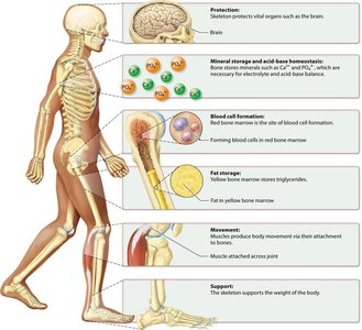
Long Bone Anatomy
Long bones, such as the femur, are composed of several distinct regions and tissues. - Diaphysis: The shaft, composed mainly of compact bone. - Epiphysis: The ends, containing spongy bone and red marrow. - Periosteum: The fibrous membrane covering the bone's exterior. - Medullary Cavity: The hollow interior filled with yellow marrow. - Endosteum: The membrane lining the medullary cavity. - Articular Cartilage: Covers joint surfaces for smooth movement. 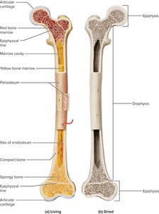
Bone Tissue Composition and Properties
Bone tissue is a specialized connective tissue with unique biomechanical properties. - Organic Matrix: Mainly collagen fibers, providing tensile strength and flexibility. - Inorganic Matrix: Hydroxyapatite crystals, conferring compressive strength and rigidity. - Composite Function: The combination allows bone to be strong, hard, and resilient. 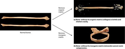
Bone Cells and Remodeling
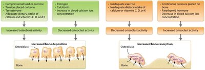
Four main cell types regulate bone formation and resorption: - Osteoprogenitor Cells: Stem cells that differentiate into osteoblasts. - Osteoblasts: Bone-forming cells that secrete osteoid and regulate mineralization. - Osteocytes: Mature bone cells maintaining the matrix and acting as mechanosensors. - Osteoclasts: Multinucleated cells that resorb bone by secreting acids and enzymes. Bone remodeling is a continuous process balancing deposition and resorption, regulated by hormones and mechanical forces. 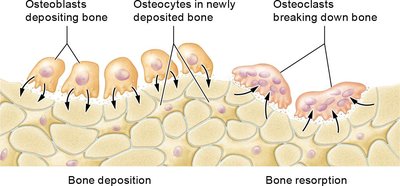
Cartilage and Its Types
Cartilage Structure and Distribution
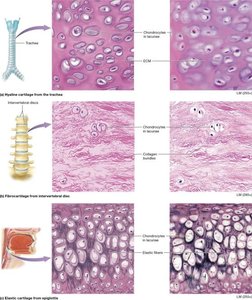
Cartilage is a resilient connective tissue formed by chondroblasts and maintained by chondrocytes in lacunae. It is found in various locations throughout the body, providing support and flexibility. - Hyaline Cartilage: Most common, found in joints, nose, trachea, and costal cartilage. - Elastic Cartilage: Contains elastic fibers, found in the ear and epiglottis. - Fibrocartilage: Contains dense collagen bundles, found in intervertebral discs and pubic symphysis. 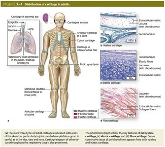
Bone Tissue Histology
Compact vs. Spongy Bone
- Compact (Cortical) Bone: Dense, organized into osteons (Haversian systems) with concentric lamellae and central canals. - Spongy (Cancellous) Bone: Lattice of trabeculae, oriented along stress lines, filled with marrow. 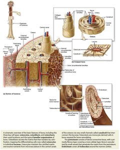
Classification of Joints
Joint Structure and Mobility
Joints are classified by their structure and degree of mobility: - Fibrous Joints: Bones united by fibrous tissue; mostly immovable (e.g., sutures, syndesmoses, gomphoses). - Cartilaginous Joints: Bones united by cartilage; slightly movable (e.g., synchondroses, symphyses). - Synovial Joints: Freely movable, with a fluid-filled cavity lined by synovial membrane; subclassified by shape (plane, hinge, pivot, condyloid, saddle, ball-and-socket). 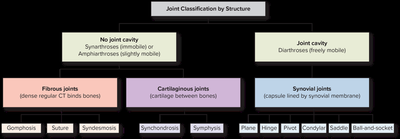
Axial and Appendicular Skeleton
Axial Skeleton
- Skull: Cranium and facial bones. - Vertebral Column: Cervical, thoracic, lumbar vertebrae, sacrum, and coccyx. - Thoracic Cage: Sternum and ribs.
Appendicular Skeleton
- Pectoral Girdle: Clavicle and scapula. - Upper Limb: Humerus, radius, ulna, carpals, metacarpals, phalanges. - Pelvic Girdle: Hip bones (ilium, ischium, pubis). - Lower Limb: Femur, patella, tibia, fibula, tarsals, metatarsals, phalanges.
Muscular System
Overview of Muscle Types
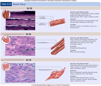
Muscle tissue is classified into three types based on structure, control, and location: - Skeletal Muscle: Voluntary, striated, attached to bones. - Cardiac Muscle: Involuntary, striated, found in the heart. - Smooth Muscle: Involuntary, non-striated, found in walls of hollow organs. 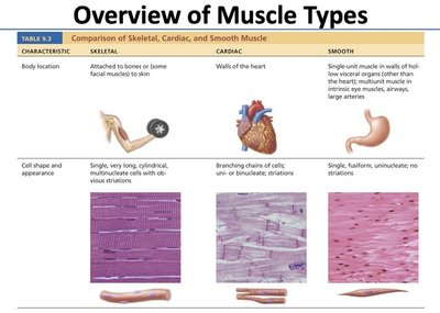
Skeletal Muscle Structure
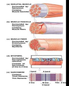
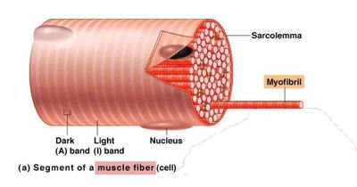
Skeletal muscle is organized hierarchically: - Whole Muscle → Fascicle → Muscle Fiber (Cell) → Myofibril → Sarcomere - Sarcomere: The contractile unit, composed of thick (myosin) and thin (actin) filaments, bounded by Z-lines. 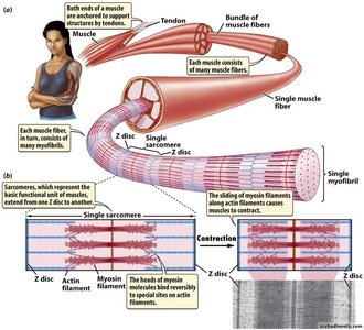
Sliding Filament Theory
Muscle contraction occurs when actin filaments slide past myosin filaments, shortening the sarcomere. - Calcium ions bind to troponin, shifting tropomyosin and exposing myosin-binding sites on actin. - ATP powers the cross-bridge cycle: attachment, power stroke, detachment, and re-cocking of myosin heads.
Neuromuscular Junction and Excitation-Contraction Coupling
The neuromuscular junction (NMJ) is the synapse between a motor neuron and a muscle fiber. - Sequence: Action potential → Ca²⁺ influx → ACh release → ACh binds nAChRs → End plate potential → Muscle action potential → Ca²⁺ release from sarcoplasmic reticulum → Contraction. - Acetylcholinesterase terminates ACh action, enabling relaxation. 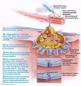
Clinical Correlations
Osteoporosis
Osteoporosis is characterized by reduced bone mass and microarchitectural deterioration, increasing fracture risk. - Pathophysiology: Estrogen deficiency increases osteoclast activity via RANKL/OPG axis. - Diagnosis: DEXA scan T-score ≤ -2.5. - Management: Bisphosphonates, denosumab, teriparatide, calcium/vitamin D supplementation, exercise, fall prevention.
Myasthenia Gravis
Myasthenia gravis is an autoimmune disorder targeting nAChRs at the NMJ, causing fatigable muscle weakness. - Diagnosis: Edrophonium test, anti-nAChR antibody titer, EMG. - Treatment: Acetylcholinesterase inhibitors, immunosuppressants, plasmapheresis, thymectomy.
Tables
Selected Major Bones — Location and Clinical Correlation
Bone | Skeleton Division | Anatomical Location | Clinical Correlation |
|---|---|---|---|
Temporal Bone | Axial (Cranium) | Lateral skull; houses middle & inner ear | Temporal bone fracture can damage facial nerve (CN VII) and cochlea |
Sternum | Axial (Thorax) | Anterior midline of chest | Site of bone marrow biopsy; CPR chest compressions compress sternum 5–6 cm |
Clavicle | Appendicular (Pectoral Girdle) | Horizontal strut between manubrium and scapula | Most commonly fractured bone in humans; falls on outstretched hand |
Humerus | Appendicular (Upper Limb) | Single bone of the arm | Radial nerve injury with spiral groove fracture causes wrist drop |
Radius | Appendicular (Upper Limb) | Lateral forearm | Colles' fracture (distal radius) — most common fracture from fall on outstretched hand |
Femur | Appendicular (Lower Limb) | Thigh — longest and strongest bone | Hip fracture (femoral neck) — major cause of morbidity/mortality in elderly with osteoporosis |
Tibia | Appendicular (Lower Limb) | Medial leg; weight-bearing bone | Stress fractures common in runners; shin splints involve periosteum |
Vertebrae (L4–L5) | Axial (Vertebral Column) | Lumbar spine | Most common site of herniated intervertebral disc; lumbar puncture at L4–L5 level |
Major Skeletal Muscles — Origin, Insertion, Action, and Innervation
Muscle | Origin | Insertion | Primary Action | Innervation |
|---|---|---|---|---|
Deltoid | Clavicle; acromion; scapular spine | Deltoid tuberosity of humerus | Arm abduction (middle fibers); flexion (anterior); extension (posterior) | Axillary nerve (C5, C6) |
Biceps Brachii | Coracoid process; supraglenoid tubercle | Radial tuberosity; bicipital aponeurosis | Forearm flexion; supination | Musculocutaneous nerve (C5, C6) |
Triceps Brachii | Infraglenoid tubercle; posterior humerus | Olecranon process of ulna | Forearm extension | Radial nerve (C6, C7, C8) |
Pectoralis Major | Clavicle; sternum; costal cartilages 1–6 | Intertubercular groove of humerus | Arm flexion, adduction, medial rotation | Medial and lateral pectoral nerves (C5–T1) |
Latissimus Dorsi | Spinous processes T7–T12; thoracolumbar fascia; iliac crest; lower ribs | Intertubercular groove of humerus | Arm extension, adduction, medial rotation | Thoracodorsal nerve (C6, C7, C8) |
Rectus Abdominis | Pubic symphysis; pubic crest | Xiphoid process; costal cartilages 5–7 | Trunk flexion; compression of abdomen | Intercostal nerves T7–T12 |
Iliopsoas (Iliacus + Psoas Major) | Iliac fossa (iliacus); lumbar vertebral bodies (psoas) | Lesser trochanter of femur | Hip flexion; thigh medial rotation | Femoral nerve (L2, L3, L4) |
Gluteus Maximus | Posterior iliac crest; sacrum; coccyx | Gluteal tuberosity; iliotibial tract | Hip extension; lateral rotation | Inferior gluteal nerve (L5, S1, S2) |
Quadriceps Femoris (group) | ASIS; anterior iliac spine; anterior femur (three vasti) | Tibial tuberosity via patellar tendon | Knee extension; hip flexion (rectus femoris) | Femoral nerve (L2, L3, L4) |
Gastrocnemius | Medial and lateral femoral condyles | Calcaneal (Achilles) tendon → calcaneus | Plantarflexion of foot; knee flexion | Tibial nerve (S1, S2) |
Key Terminology
- Osteoblast: Bone-forming cell that synthesizes and mineralizes osteoid. - Osteoclast: Bone-resorbing cell that dissolves mineral and degrades matrix. - Osteocyte: Mature bone cell in lacunae, mechanosensor. - Periosteum: Membrane covering bone, essential for growth and repair. - Synovial Joint: Freely movable joint with a cavity lined by synovial membrane. - Osteoporosis: Disease with reduced bone density and increased fracture risk. - Sarcomere: Contractile unit of muscle, site of cross-bridge cycling. - Sliding Filament Theory: Actin slides past myosin, shortening sarcomere. - Neuromuscular Junction (NMJ): Synapse between motor neuron and muscle fiber. - Motor Unit: Motor neuron and all muscle fibers it innervates. - Acetylcholinesterase: Enzyme terminating ACh action at NMJ. - Myasthenia Gravis: Autoimmune disorder affecting NMJ.
Additional info:
- The notes expand on brief points with academic context, definitions, and examples. - Images included are directly relevant to the adjacent explanations and reinforce understanding. - Tables are recreated to preserve clinical correlations and muscle anatomy. ----------------------------------------
