 Indietro
Study Guide: Skeletal System – Structure, Function, and Regional Anatomy
Guida di studio - Note intelligenti
 Appunti personalizzati basati sui tuoi materiali, ampliati con definizioni chiave, esempi e contesto.
Skeletal System Overview
Structure and Function
The skeletal system is a complex framework composed of bones, cartilages, joints, and ligaments. It provides structural support, protects vital organs, facilitates movement, and serves as a reservoir for minerals.
Bones: Provide rigidity and structure; most of the skeleton is bone.
Cartilage: Occurs in isolated areas, offering flexibility and cushioning.
Ligaments: Connect bones and reinforce joints.
Joints: Allow movement between bones.
Body Mass: The skeleton accounts for approximately 20% of body mass.
Major Divisions of the Skeleton
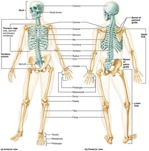
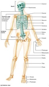
The skeleton is divided into two major subdivisions: the axial and appendicular skeletons.
Axial Skeleton: Consists of 80 bones in three regions: skull, vertebral column, and thoracic cage. Functions include forming the longitudinal axis, supporting the head, neck, and trunk, and protecting the brain, spinal cord, and thoracic organs.
Appendicular Skeleton: Composed of the bones of the limbs and their girdles (pectoral and pelvic), facilitating movement and interaction with the environment.
Axial Skeleton
Skull: Cranial and Facial Bones
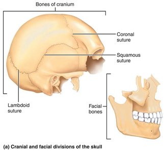
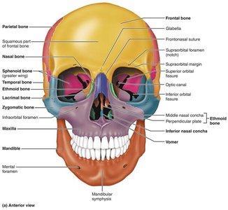
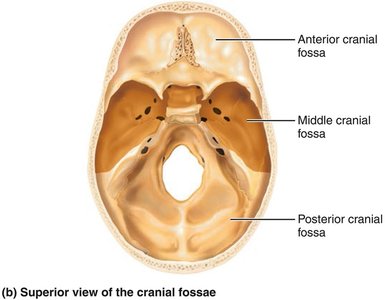
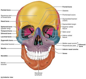
The skull is formed by two sets of bones: cranial bones (cranium) and facial bones. Cranial bones enclose the brain, while facial bones form the framework of the face and provide openings for air and food passage.
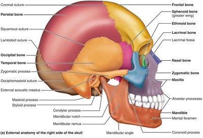
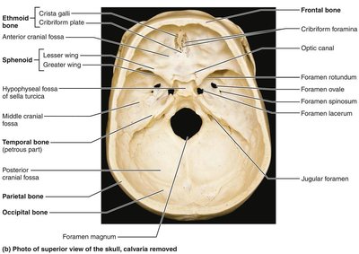
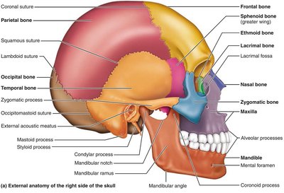
Cranial Bones: Frontal (1), Parietal (2), Temporal (2), Occipital (1), Ethmoid (1), Sphenoid (1).
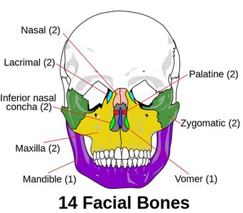
Facial Bones: Mandible (1), Maxillary (2), Zygomatic (2), Nasal (2), Lacrimal (2), Palatine (2), Vomer (1), Inferior nasal conchae (2).
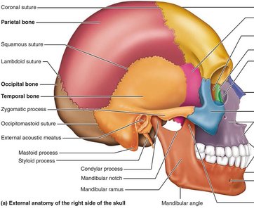
Sutures: Joints between skull bones, with a serrated appearance (e.g., coronal, sagittal, lambdoid, squamous).
Key Cranial Bone Features
Frontal Bone: Forms the forehead and anterior cranium; contains frontal sinuses.
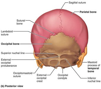
Parietal Bones: Form superior and lateral aspects; four sutures mark their articulations.
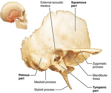
Temporal Bones: Inferolateral aspects; include zygomatic process, tympanic region, mastoid process, and house ear cavities.
Occipital Bone: Posterior wall; contains foramen magnum and occipital condyles.
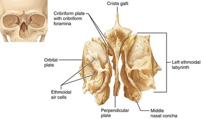
Ethmoid Bone: Deepest skull bone; forms roof of nasal cavity and medial wall of orbits.
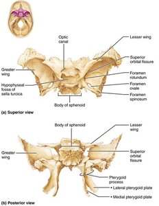
Sphenoid Bone: Bat-shaped, keystone bone; articulates with all other cranial bones.
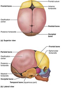
Infant Skull
Infant skull has more bones than adult skull; bones are connected by fontanelles (unossified fibrous membranes) to ease birth and allow brain growth.
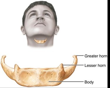
Hyoid Bone
Not a bone of the skull; only bone not articulating directly with another bone.
Anchored by ligaments; serves as a base for the tongue and attachment site for muscles of swallowing and speech.
Facial Bones
The facial skeleton consists of 14 bones, most of which are paired. These bones form the structure of the face, house the teeth, and provide attachment sites for facial muscles.
Mandible: Largest, strongest bone; contains sockets for teeth.
Maxillary Bones: Form upper jaw; hold upper teeth; contain maxillary sinuses.
Zygomatic Bones: Form cheekbones.
Nasal Bones: Form bridge of nose.
Lacrimal Bones: Medial walls of orbits; house lacrimal sac for tear drainage.
Palatine Bones: Form plates of palate.
Vomer: Forms part of nasal septum.
Inferior Nasal Conchae: Form part of lateral walls of nasal cavity.
Orbits and Paranasal Sinuses
Orbits: Cavities encasing eyes and lacrimal glands; formed by parts of seven bones.
Paranasal Sinuses: Mucosa-lined, air-filled spaces in five skull bones; functions include warming/humidifying air, lightening the skull, and enhancing voice resonance.
Vertebral Column
Structure and Regions
The vertebral column, or spine, extends from the skull to the pelvis and consists of 26 irregular bones in five major regions. It transmits weight, protects the spinal cord, and provides attachment points for ribs and muscles.
Cervical: 7 vertebrae
Thoracic: 12 vertebrae
Lumbar: 5 vertebrae
Sacrum: 1 bone (fusion of several vertebrae)
Coccyx: 1 bone (fusion of several vertebrae)
Curvatures
Cervical and Lumbar: Concave posteriorly
Thoracic and Sacral: Convex posteriorly
Curvatures increase resilience and flexibility.
Abnormal Curvatures
Scoliosis: Abnormal lateral rotation; may cause breathing difficulties.
Kyphosis: Abnormal dorsal thoracic curvature (hunchback).
Lordosis: Accentuated lumbar curvature (swayback).
Intervertebral Discs
Nucleus Pulposus: Inner gelatinous core; provides elasticity and compressibility.
Anulus Fibrosus: Outer collar of collagen and fibrocartilage; limits expansion.
Herniated Disc: Rupture of anulus fibrosus, protrusion of nucleus pulposus; may press on nerves.
Vertebral Structure
Body (Centrum): Anterior weight-bearing region.
Vertebral Foramen: Enclosure formed by body and arch.
Processes: Spinous, transverse, superior/inferior articular.
Special Vertebrae
Atlas (C1): No body or spinous process; articulates with occipital condyles.
Axis (C2): Has dens; pivot for rotation.
C7: Vertebra prominens; landmark.
Thoracic Cage
Structure and Function
The thoracic cage is composed of thoracic vertebrae, ribs, sternum, and costal cartilages. It protects vital organs, supports shoulder girdles and upper limbs, and provides muscle attachment sites.
Sternum: Manubrium, body, xiphoid process.
Ribs: 12 pairs; true ribs (1–7), false ribs (8–10), floating ribs (11–12).
Appendicular Skeleton
Pectoral Girdle and Upper Extremities
The pectoral girdle consists of clavicles and scapulae, attaching upper limbs to the axial skeleton and providing mobility.
Clavicle: S-shaped; articulates with sternum and scapula.
Scapula: Triangular; features acromion, important for IM injections.
Upper Limb Bones: Humerus (arm), radius and ulna (forearm), carpals (wrist), metacarpals (palm), phalanges (fingers).
Pelvic Girdle and Lower Extremities
The pelvic girdle is formed by two hip bones and the sacrum, attaching lower limbs to the axial skeleton and supporting pelvic organs.
Coxal Bone: Formed by fusion of ilium, ischium, and pubis.
Pelvis: Includes hip bones, sacrum, coccyx; female pelvis is wider and adapted for childbirth.
Lower Limb Bones: Femur (thigh), tibia and fibula (leg), tarsals (foot), metatarsals, phalanges (toes).
Key Points for Lower Extremity
Femur: Largest and strongest bone; articulates with acetabulum, tibia, and patella.
Tibia: Medial, weight-bearing bone.
Fibula: Lateral, not weight-bearing.
Foot: Tarsus (7 bones), metatarsals (5), phalanges (14).
Summary Table: Major Bones of the Human Skeleton
Region | Main Bones |
|---|---|
Skull | Frontal, Parietal, Temporal, Occipital, Ethmoid, Sphenoid, Mandible, Maxilla, Zygomatic, Nasal, Lacrimal, Palatine, Vomer, Inferior nasal conchae |
Vertebral Column | Cervical (7), Thoracic (12), Lumbar (5), Sacrum, Coccyx |
Thoracic Cage | Sternum, Ribs (12 pairs) |
Pectoral Girdle | Clavicle, Scapula |
Upper Limb | Humerus, Radius, Ulna, Carpals, Metacarpals, Phalanges |
Pelvic Girdle | Ilium, Ischium, Pubis |
Lower Limb | Femur, Tibia, Fibula, Tarsals, Metatarsals, Phalanges |
Additional info: The notes above expand on brief points with academic context, definitions, and examples to ensure completeness and clarity for exam preparation.
