 Indietro
Study Guide: Synovial Joints and Joint Disorders (Anatomy & Physiology, Chapter 8)
Guida di studio - Note intelligenti
 Appunti personalizzati basati sui tuoi materiali, ampliati con definizioni chiave, esempi e contesto.
Joints: Synovial Joint Structure and Function
Overview of Synovial Joints
Synovial joints are the most common and movable type of joint in the human body. They possess unique structural features that allow for a wide range of motion but also introduce certain weaknesses. - Definition: Synovial joints are freely movable joints characterized by the presence of a synovial cavity filled with synovial fluid. - Main Synovial Joints: Jaw (temporomandibular), shoulder, elbow, hip, and knee. - General Features: All synovial joints have articular cartilage, a joint cavity, an articular capsule, synovial fluid, reinforcing ligaments, and nerves and blood vessels.
Shoulder (Glenohumeral) Joint
Anatomy and Function
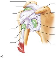
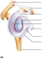
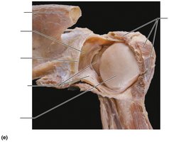
The shoulder joint is the most freely moving joint in the body, sacrificing stability for mobility. It is a ball-and-socket joint formed by the head of the humerus and the glenoid cavity of the scapula. - Ball-and-Socket Structure: The large, hemispherical head of the humerus fits into the small, shallow glenoid cavity, similar to a golf ball on a tee. - Articular Capsule: Thin and loose, allowing for extensive movement. - Reinforcing Structures: Muscle tendons, especially the tendon of the long head of the biceps brachii, provide stability. The rotator cuff (subscapularis, supraspinatus, infraspinatus, teres minor) encircles the joint. 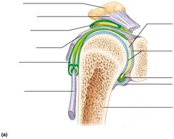
Elbow Joint
Anatomy and Function
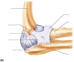
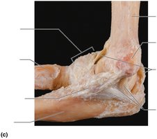
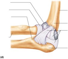
The elbow joint is a hinge joint formed by the humerus, ulna, and radius. It allows for flexion and extension of the forearm. - Articular Capsule: Encloses the joint and contains synovial fluid. - Ligaments: The ulnar collateral, radial collateral, and anular ligaments reinforce the joint. - Muscle Attachments: Tendons of the brachialis and triceps muscles contribute to movement and stability. 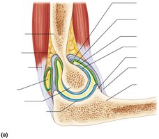
Knee Joint
Anatomy and Function
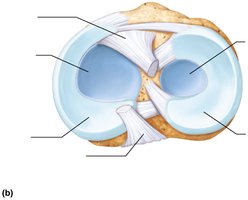
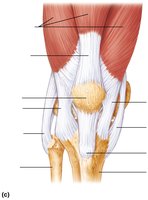
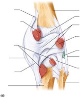
The knee is the largest and most complex joint in the body, consisting of three joints surrounded by a single cavity. - Femoropatellar Joint: Plane joint allowing gliding motion during knee flexion. - Tibiofemoral Joint: Hinge joint between femoral condyles and menisci of tibia, allowing flexion, extension, and some rotation. - Ligaments and Menisci: Cruciate ligaments (anterior and posterior), collateral ligaments (fibular and tibial), and menisci (lateral and medial) stabilize the joint. 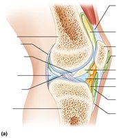
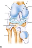
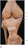
The "Unhappy Triad" Injury
The "unhappy triad" refers to a simultaneous injury of the anterior cruciate ligament (ACL), tibial collateral ligament, and medial meniscus, often caused by a lateral blow to the knee. 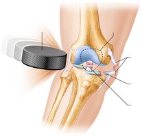
Disorders of Joints
Common Joint Injuries
- Cartilage Tears: Caused by compression and shear stress; fragments may cause joint locking. Cartilage rarely repairs itself and may require arthroscopic surgery. 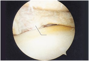 - Sprains: Stretching or tearing of ligaments, commonly in the ankle, knee, and lumbar region. Partial tears heal slowly; complete tears may require surgical intervention. - Dislocations (Luxations): Bones forced out of alignment, often accompanied by sprains and inflammation. Subluxation is a partial dislocation.
Inflammatory and Degenerative Conditions
Bursitis and Tendonitis
- Bursitis: Inflammation of a bursa, usually caused by trauma or friction. - Tendonitis: Inflammation of tendon sheaths, typically due to overuse. Both conditions are treated with rest, ice, and anti-inflammatory drugs.
Arthritis
Arthritis encompasses over 100 types of joint diseases, characterized by pain, stiffness, and swelling. Acute forms are often bacterial and treated with antibiotics. Chronic forms include osteoarthritis, rheumatoid arthritis, and gouty arthritis.
Osteoarthritis (OA)
- Most common, irreversible, degenerative arthritis. - Cartilage breakdown exceeds repair, leading to bone spurs (osteophytes). - Symptoms include joint stiffness and crepitus. - Treatment: moderate activity, mild pain relievers, capsaicin creams.
Rheumatoid Arthritis (RA)
- Chronic, inflammatory, autoimmune disease. - Immune system attacks synovial membrane, leading to synovitis, pannus formation, cartilage erosion, and ankylosis. - Symptoms: joint pain, swelling, anemia, osteoporosis, muscle weakness, cardiovascular issues. - Treatment: anti-inflammatory drugs, immune suppressants, joint replacement. 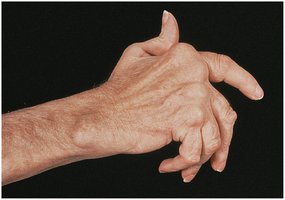
Gouty Arthritis
- Caused by deposition of uric acid crystals in joints, leading to inflammation. - More common in men; often affects the base of the great toe. - Untreated cases may result in joint fusion. - Treatment: drugs, hydration, avoidance of alcohol and purine-rich foods.
Lyme Disease
- Caused by bacteria transmitted by tick bites. - Symptoms: skin rash, flu-like symptoms, foggy thinking, joint pain, and arthritis. - Treatment: long course of antibiotics.
Summary Table: Major Synovial Joints and Their Features
Joint | Type | Main Features | Common Injuries |
|---|---|---|---|
Shoulder (Glenohumeral) | Ball-and-socket | High mobility, rotator cuff, thin capsule | Dislocation, rotator cuff tear |
Elbow | Hinge | Strong ligaments, flexion/extension | Sprain, dislocation |
Knee | Hinge/plane | Menisci, cruciate/collateral ligaments | "Unhappy triad", meniscus tear |
Key Terms and Definitions
Synovial fluid: Lubricating fluid within the joint cavity.
Articular cartilage: Hyaline cartilage covering bone surfaces in joints.
Bursa: Fluid-filled sac reducing friction between tissues.
Ligament: Connective tissue connecting bones.
Meniscus: Crescent-shaped cartilage in the knee.
Pannus: Abnormal tissue in RA that erodes cartilage.
Ankylosis: Joint immobilization due to scar tissue.
Equations and Biomechanics
Joint Stability Equation
Joint stability is influenced by the shape of articular surfaces, ligament strength, and muscle tone.
Conclusion
Understanding the anatomy and physiology of synovial joints is essential for recognizing their function, vulnerabilities, and common disorders. Proper care and early intervention can prevent or mitigate joint injuries and degenerative conditions.
