 Indietro
Study Guide: The Appendicular Skeleton (Anatomy & Physiology)
Guida di studio - Note intelligenti
 Appunti personalizzati basati sui tuoi materiali, ampliati con definizioni chiave, esempi e contesto.
The Appendicular Skeleton
Overview and Functions
The appendicular skeleton consists of the bones of the limbs and the supporting girdles that connect them to the trunk. It plays a crucial role in movement and weight support, providing attachment points for muscles and facilitating locomotion and manipulation.
Attachment points: Muscles involved in walking, hand movements, and other activities attach to these bones.
Weight support: Lower limbs support the body's weight during standing and movement.
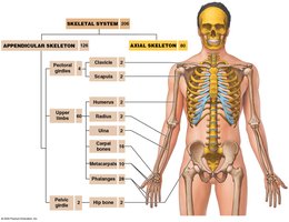
Pectoral Girdle
Bones, Functions, and Surface Features
The pectoral (shoulder) girdle connects each arm to the trunk and provides a basis for arm movement. Each girdle consists of one clavicle and one scapula.
Clavicle: S-shaped bone, connects with the axial skeleton at the manubrium (sternal end) and articulates with the acromion of the scapula (acromial end).
Scapula: Flat, triangular bone located on the posterior side of the rib cage. Articulates with the clavicle and humerus.
Mobility: The shoulder is highly mobile but not very strong due to its skeletal muscle support.
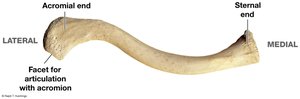
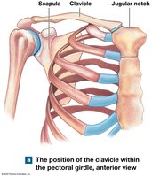
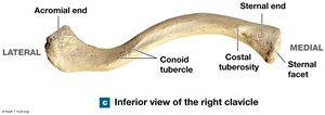
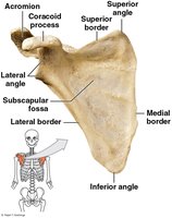
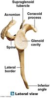
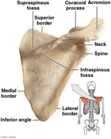
Scapula Surface Features
Borders: Superior, medial (vertebral), and lateral (axillary).
Angles: Superior, inferior, and lateral.
Processes: Coracoid (smaller, anterior), acromion (larger, posterior), and spine (ridge dividing the posterior surface).
Fossae: Subscapular (anterior), supraspinous (above spine), infraspinous (below spine).
Glenoid cavity: Cup-shaped surface at the lateral angle, articulates with the humerus.
Upper Limbs
Bones, Functions, and Surface Features
The skeleton of the upper limbs includes the bones of the arm, forearm, wrist, and hand. These bones facilitate a wide range of movements and manipulations.
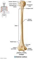
Arm: Humerus (shoulder to elbow)
Forearm: Ulna (medial), radius (lateral)
Wrist: Eight carpal bones
Hand: Five metacarpals, phalanges (fingers)
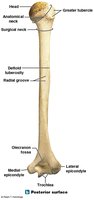
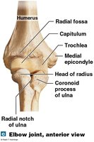
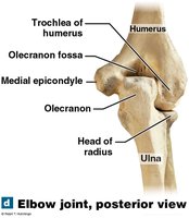
Forearm Bones
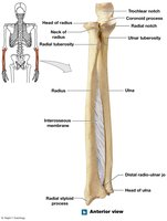
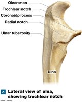
Ulna: Medial bone, olecranon forms the elbow point, trochlear notch articulates with humerus, coronoid process fits into coronoid fossa during flexion.
Radius: Lateral bone, head articulates with capitulum of humerus, radial tuberosity is attachment for biceps brachii.
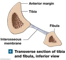
Interosseous membrane: Fibrous sheet connecting ulna and radius.
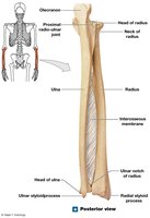
Wrist and Hand Bones
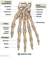
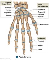
Carpals: Eight bones, four proximal (scaphoid, lunate, triquetrum, pisiform), four distal (trapezium, trapezoid, capitate, hamate).
Metacarpals: Five long bones, numbered from lateral to medial.
Phalanges: Pollex (thumb) has two, other fingers have three each.
Pelvic Girdle
Bones, Functions, and Surface Features
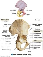
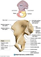
The pelvic girdle attaches the lower limb to the axial skeleton and is strong to bear body weight and provide mobility. It consists of two hip bones (coxal bones), each formed by the fusion of the ilium, ischium, and pubis.
Ilium: Superior portion, provides attachment points for muscles and ligaments.
Ischium: Posterior and inferior, supports body weight when sitting.
Pubis: Anterior and inferior, forms the pubic symphysis.
Acetabulum: Socket for the head of the femur.
Obturator foramen: Space for muscle attachment.
Pelvis Structure
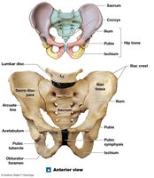
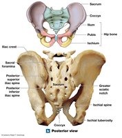
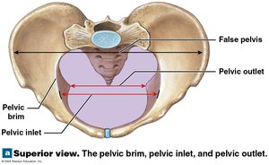
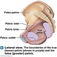
Pelvic inlet: Space enclosed by the pelvic brim.
True (lesser) pelvis: Inferior to the pelvic brim.
False (greater) pelvis: Superior to the pelvic brim.
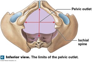
Pelvic outlet: Opening bounded by coccyx, ischial tuberosities, and pubic symphysis.
Perineum: Surface region bordered by inferior edges of pelvis.
Sex Differences in the Pelvis
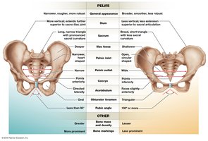
Female pelvis: Broader, smoother, lighter, wider pelvic outlet, triangular obturator foramen, pubic angle ≥ 100°.
Male pelvis: Narrower, rougher, more robust, oval obturator foramen, pubic angle ≤ 90°.
Lower Limbs
Bones, Functions, and Surface Features
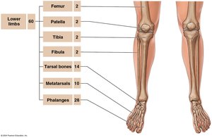
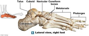
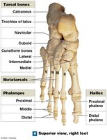
The lower limb consists of the femur, patella, tibia, fibula, tarsal bones, metatarsals, and phalanges. These bones are specialized for weight bearing and movement.
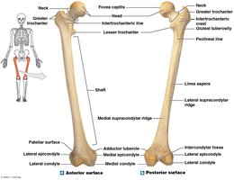
Femur: Longest, heaviest bone; articulates with hip and tibia.
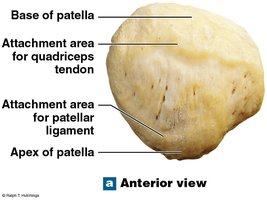
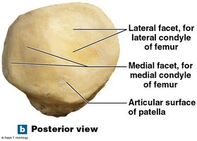
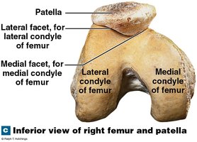
Patella: Sesamoid bone within quadriceps tendon.
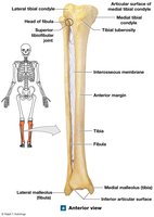
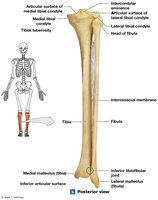
Tibia: Medial, weight-bearing bone.
Fibula: Lateral, stabilizes ankle.
Tarsals: Seven bones, including talus and calcaneus.
Metatarsals: Five long bones.
Phalanges: Four toes have three each, hallux has two.
Arches of the Foot
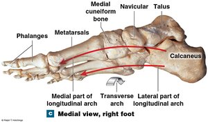
Longitudinal arch: Ties calcaneus to distal metatarsals, keeps medial plantar surface elevated.
Transverse arch: Curvature from one side of the foot to the other.
Flatfeet: Condition where normal arches are lost or never form.
Individual Skeletal Variation
Sex Differences and Age-Related Changes
Skeletal features can reveal muscle strength, medical history, sex, age, and body size. Modifications in the female pelvis accommodate childbearing, and hormone relaxin increases pelvic inlet and outlet size during pregnancy.
Table: Sex Differences in the Human Skeleton
Feature | Male | Female |
|---|---|---|
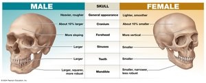
Skull | Heavier, rougher, larger, more sloping forehead | Lighter, smoother, smaller, more vertical forehead |
Pelvis | Narrower, rougher, more robust, pubic angle < 90° | Broader, smoother, less robust, pubic angle > 100° |
Other | Greater bone mass and density | Less bone mass and density |
Table: Age-Related Changes in the Skeleton
Change | Description |
|---|---|
Bone mass | Decreases with age |
Bone fusion | Epiphyseal plates fuse in adulthood |
Joint changes | Cartilage wears down, leading to arthritis |
Fracture healing | Slower in older adults |
Additional info: Age-related changes include osteoporosis, increased fracture risk, and decreased flexibility due to loss of bone and cartilage.
