 Indietro
Study Guide: The Appendicular Skeleton (Bones, Part 2)
Guida di studio - Note intelligenti
 Appunti personalizzati basati sui tuoi materiali, ampliati con definizioni chiave, esempi e contesto.
The Appendicular Skeleton
Overview
The appendicular skeleton consists of the bones that support the limbs and enable movement. It includes the pectoral girdle, pelvic girdle, and the bones of the upper and lower limbs. This system is essential for mobility and manipulation of the environment.
Pectoral girdle: Attaches the upper limbs to the trunk.
Pelvic girdle: Attaches the lower limbs to the trunk.
Upper and lower limbs share a similar structural plan but differ in function.
Enables a wide variety of movements such as walking, throwing, and writing.
The Pectoral Girdle
Structure and Function
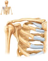
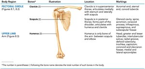
The pectoral girdle is composed of the clavicle and scapula. It provides attachment for muscles that move the upper limb and is designed for mobility rather than stability.
Clavicle: S-shaped bone articulating medially with the manubrium and laterally with the scapula.
Scapula: Triangular bone with extensive muscular attachments; does not join the axial skeleton directly.
Only the clavicle articulates with the axial skeleton, allowing the scapula to move freely.
The glenoid cavity is shallow, favoring flexibility over stability.
Clavicle
The clavicle is a slender, S-shaped bone that extends horizontally across the superior thorax. It serves as a strut to keep the scapula and arms laterally positioned and transmits compression forces from the upper limbs to the axial skeleton.
Sternal end: Articulates with the manubrium.
Acromial end: Articulates with the scapula.
Provides attachment for muscles.
Scapula
The scapula is a thin, triangular bone located on the dorsal surface of the rib cage, between ribs 2–7. It has three borders (superior, medial, lateral) and three angles (lateral, superior, inferior). The glenoid cavity at the lateral angle articulates with the humerus, forming the shoulder joint.
Major landmarks: Acromion, coracoid process, spine, suprascapular notch, subscapular fossa.
Provides attachment for muscles and forms part of the shoulder joint.
The Upper Limb
Arm
The arm is the region between the shoulder and elbow, containing the humerus, which is the only bone of the arm and the longest and strongest bone of the upper limb.
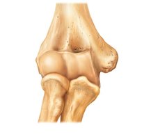
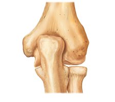
Humerus: Articulates with the scapula at the shoulder and with the radius and ulna at the elbow.
Features include the head, anatomical neck, greater and lesser tubercles, deltoid tuberosity, trochlea, capitulum, epicondyles, and fossae.
Forearm
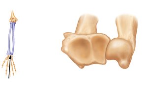
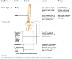
The forearm is formed from the radius and ulna. These bones articulate with each other at the proximal and distal radioulnar joints and are interconnected by the interosseous membrane.
Ulna: Main bone forming the elbow joint with the humerus; plays little role in hand movement.
Radius: Contributes heavily to the wrist joint; when the radius moves, the hand moves with it.
In anatomical position, the radius is lateral and the ulna is medial.
Hand
The hand consists of the carpus (wrist), metacarpus (palm), and phalanges (fingers). Each finger has three phalanges except the thumb, which has two.
Carpals: Eight marble-sized bones arranged in two rows.
Metacarpals: Five bones forming the palm, numbered from thumb to little finger.
Phalanges: Fourteen bones forming the fingers.
Summary Table: Bones of the Upper Limb
This table summarizes the bones, their locations, and key markings in the upper limb.
Body Region | Bones | Location | Markings |
|---|---|---|---|
Pectoral Girdle | Clavicle, Scapula | Superior thorax, dorsal rib cage | Sternal/acromial ends, glenoid cavity, acromion, coracoid process |
Arm | Humerus | Shoulder to elbow | Head, tubercles, epicondyles, trochlea, capitulum |
Forearm | Ulna, Radius | Elbow to wrist | Olecranon, styloid processes, radial tuberosity |
Hand | Carpals, Metacarpals, Phalanges | Wrist, palm, fingers | Proximal/distal rows, bases, heads |
The Pelvic Girdle
Structure and Function
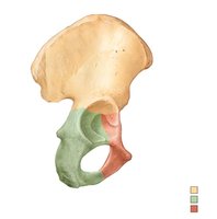
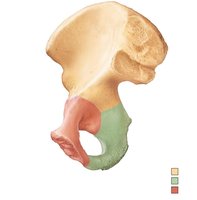
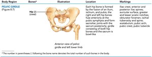
The pelvic girdle attaches the lower limbs to the spine and supports visceral organs. It consists of paired hip bones (coxal bones) and the sacrum. The acetabulum is a deep socket that secures the head of the femur.
Ilium: Superior region, site of muscle attachment.
Ischium: Posteroinferior region, strongest part of the hip bone.
Pubis: Anterior region, joined at the pubic symphysis.
Pelvic Structure and Childbearing
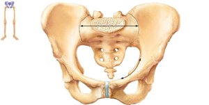
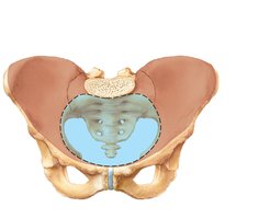
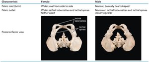
The bony pelvis is divided into the false (greater) pelvis and true (lesser) pelvis, separated by the pelvic brim. The female pelvis is wider, shallower, and lighter, adapted for childbearing.
Pelvic inlet: Largest diameter right to left.
Pelvic outlet: Largest diameter anteroposteriorly.
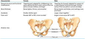
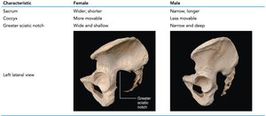
Major differences between male and female pelves are related to childbearing.
Comparison Table: Male vs. Female Pelvis
This table compares the structural differences between male and female pelves.
Characteristic | Female | Male |
|---|---|---|
General structure | Lighter, wider, shallower | Heavier, narrower, deeper |
Pelvic inlet | Wider, oval | Narrow, heart-shaped |
Pelvic outlet | Wider | Narrower |
Pubic arch | Broader (80–90°) | Narrower (50–60°) |
The Lower Limb
Thigh
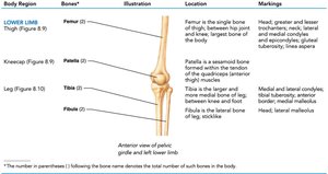
The thigh is the region between the hip and knee, containing the femur, which is the longest and strongest bone in the body. The femur's head articulates with the acetabulum.
Femur: Features include head, neck, trochanters, condyles, epicondyles, and linea aspera.
Patella: Triangular sesamoid bone embedded in the quadriceps tendon.
Leg
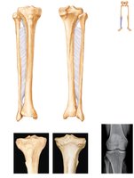
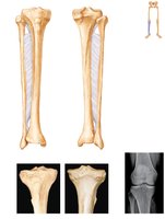
The leg is the region between the knee and ankle, composed of the tibia and fibula. The tibia receives the body's weight from the femur, while the fibula stabilizes the ankle joint.
Tibia: Medial, more massive bone; forms knee and ankle joints.
Fibula: Lateral, sticklike bone; does not contribute to the knee joint.
Interosseous membrane connects tibia and fibula.
Foot
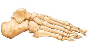
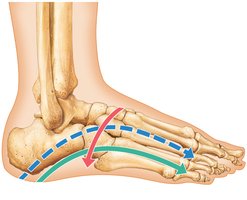
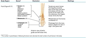
The foot is composed of the tarsus, metatarsus, and phalanges. It has three arches (medial longitudinal, lateral longitudinal, transverse) that distribute body weight and provide resilience.
Tarsus: Seven bones; body weight is borne by the talus and calcaneus.
Metatarsus: Five metatarsals; first metatarsal forms the "ball" of the foot.
Phalanges: Fourteen bones; each toe has three phalanges except the great toe, which has two.
Arches of the Foot
The foot's arches are maintained by the interlocking shapes of tarsals, ligaments, and tendons. The talus is the keystone of the medial longitudinal arch, and the cuboid is the keystone of the lateral longitudinal arch.
Arches distribute body weight and provide resilience during movement.
Summary Table: Bones of the Lower Limb
This table summarizes the bones, their locations, and key markings in the lower limb.
Body Region | Bones | Location | Markings |
|---|---|---|---|
Pelvic Girdle | Hip (Ilium, Ischium, Pubis) | Pelvic region | Iliac crest, acetabulum, pubic arch, ischial tuberosity |
Thigh | Femur | Hip to knee | Head, neck, trochanters, condyles, linea aspera |
Leg | Tibia, Fibula | Knee to ankle | Condyles, malleoli, tuberosity |
Foot | Tarsals, Metatarsals, Phalanges | Ankle, foot, toes | Talus, calcaneus, metatarsal heads |
Additional info:
The appendicular skeleton is crucial for locomotion and manipulation, with structural adaptations for both stability and flexibility.
Mnemonic for carpal bones: "Sally left the party to take Carmen home" (Scaphoid, Lunate, Triquetrum, Pisiform, Trapezium, Trapezoid, Capitate, Hamate).
Pelvic differences are important in forensic identification and obstetrics.
