 Indietro
Study Guide: The Axial and Appendicular Skeleton
Guida di studio - Note intelligenti
 Appunti personalizzati basati sui tuoi materiali, ampliati con definizioni chiave, esempi e contesto.
The Human Skeleton: Overview
Divisions and Functions
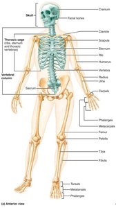
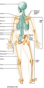
The human skeleton is a structural framework composed of bones, cartilages, joints, and ligaments. It is divided into two major regions: the axial skeleton and the appendicular skeleton. The skeleton supports the body, protects vital organs, and enables movement.
Axial Skeleton: Consists of 80 bones, including the skull, vertebral column, and thoracic cage. Forms the longitudinal axis of the body.
Appendicular Skeleton: Includes bones of the limbs and girdles that attach them to the axial skeleton.
Functions: Support, protection, movement, and mineral storage.
The Axial Skeleton
Skull: Structure and Function
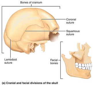
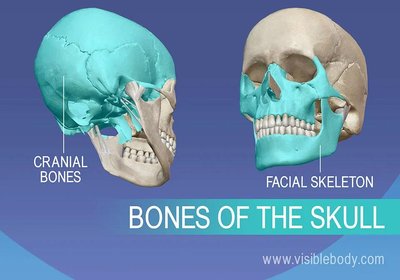
The skull is composed of cranial and facial bones, which protect the brain and form the framework of the face.
Cranial Bones: Enclose the brain and provide attachment sites for head and neck muscles.
Facial Bones: Form the face, house sensory organ cavities, provide openings for air and food, secure teeth, and anchor facial muscles.
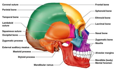
Sutures: Immovable joints connecting skull bones, with a serrated appearance.
Skull Geography
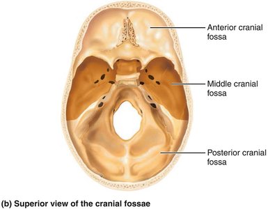
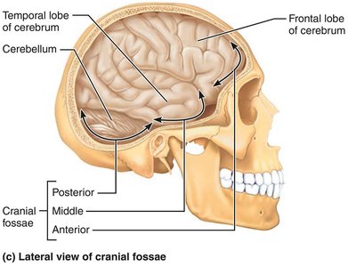
The cranium is divided into a vault and a base. The cranial vault forms the superior, lateral, and posterior portions, while the base forms the inferior aspect. Internally, the base is divided into three fossae: anterior, middle, and posterior, which house the brain.
Cranial Cavity: Encloses the brain.
Other Cavities: Middle and internal ear, nasal cavity, orbits, and sinuses.
Openings: Foramina, canals, and fissures for passage of nerves and blood vessels.
Skull Bones and Markings
Foramina: Openings for blood vessels and nerves.
Processes: Projections for muscle attachment.
Sutures: Immovable joints connecting skull bones.
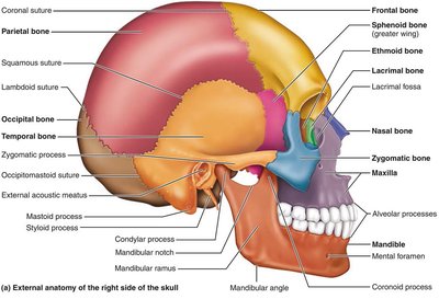
Cranial Bones
The cranium consists of eight bones: frontal, parietal (2), occipital, temporal (2), sphenoid, and ethmoid. Each bone has unique features and articulations.
Frontal Bone: Forms the forehead, superior wall of orbits, and anterior cranial fossa. Contains the glabella and frontal sinuses.
Parietal Bones: Form most of the superior and lateral aspects of the cranial vault. Articulated by coronal, sagittal, lambdoid, and squamous sutures.
Occipital Bone: Forms the posterior wall and cranial fossa. Features include the foramen magnum, occipital condyles, and nuchal lines.
Temporal Bones: Make up the inferolateral aspects and parts of the cranial base. Include squamous, tympanic, and petrous parts, with important foramina and processes.
Sphenoid Bone: Keystone bone articulating with all other cranial bones. Contains sphenoidal sinuses and sella turcica.
Ethmoid Bone: Deepest skull bone, forms part of the nasal cavity and orbits.
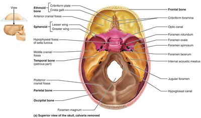
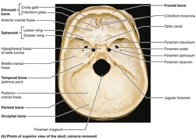
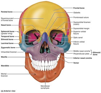
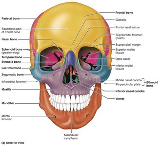
Facial Bones
The facial skeleton consists of 14 bones, including the mandible, maxillae, zygomatic, nasal, lacrimal, palatine, vomer, and inferior nasal conchae.
Mandible: Largest, strongest bone of the face; forms the lower jaw.
Maxillae: Form the upper jaw and central facial skeleton.
Zygomatic Bones: Form the cheekbones.
Nasal Bones: Form the bridge of the nose.
Lacrimal Bones: Form the medial walls of the orbits.
Palatine Bones: Complete the hard palate and form parts of the nasal cavity and orbits.
Vomer: Forms part of the nasal septum.
Inferior Nasal Conchae: Form part of the lateral walls of the nasal cavity.
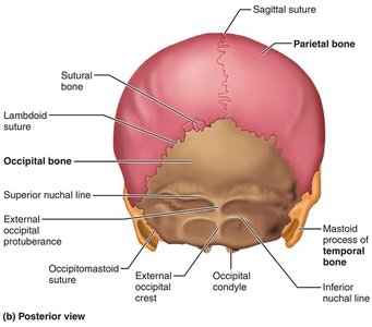
Bony Boundaries of the Orbits and Nasal Cavity
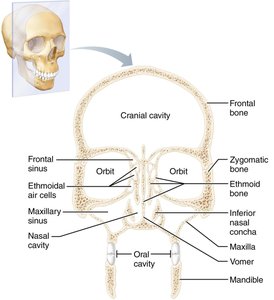
The orbits and nasal cavity are formed by multiple bones, providing protection and housing for sensory organs.
Orbits: Roof (frontal, sphenoid), lateral wall (zygomatic, sphenoid), floor (maxilla, zygomatic, palatine), medial wall (ethmoid, lacrimal, maxilla, sphenoid).
Nasal Cavity: Roof (nasal, frontal, ethmoid, sphenoid), floor (maxilla, palatine), lateral walls (maxilla, ethmoid, inferior nasal conchae, palatine), medial wall (ethmoid, vomer).
Paranasal Sinuses: Frontal, ethmoidal, sphenoidal, and maxillary sinuses lighten the skull and amplify voice.
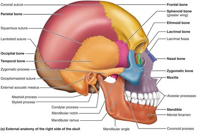
The Vertebral Column
Structure and Regions
The vertebral column extends from the skull to the pelvis, transmitting weight and protecting the spinal cord. It consists of 26 vertebrae in five regions: cervical (7), thoracic (12), lumbar (5), sacrum (1, fused), and coccyx (1, fused).
Curvatures: Cervical and lumbar (convex anteriorly), thoracic and sacral (concave anteriorly).
Functions: Shock absorption, flexibility, balance, and protection.
Structure of a Typical Vertebra
Body: Main weight-bearing structure.
Vertebral Arch: Formed by pedicles and laminae.
Processes: Spinous, transverse, superior and inferior articular processes.
Regional Features
Cervical: Smallest, with transverse foramina and bifid spinous processes.
Thoracic: Articular facets for ribs, heart-shaped body, long spinous processes.
Lumbar: Largest, kidney-shaped body, short blunt spinous processes.
The Thoracic Cage
Bones and Functions
The thoracic cage protects vital organs and supports the shoulder girdle. It consists of the sternum, ribs, and thoracic vertebrae.
Sternum: Manubrium, body, xiphoid process.
Ribs: True ribs (1-7, direct attachment), false ribs (8-12, indirect or no attachment), floating ribs (11-12).
Functions: Protection, support, respiration.
The Appendicular Skeleton
Pectoral Girdle and Upper Limb
Pectoral Girdle: Clavicle and scapula allow free movement of the upper limb.
Upper Limb: Humerus (arm), radius and ulna (forearm), carpals (wrist), metacarpals (palm), phalanges (fingers).
Pelvic Girdle and Lower Limb
Pelvic Girdle: Ilium, ischium, and pubis form the hip bone. The pelvis supports body weight and protects organs.
Lower Limb: Femur (thigh), patella (knee cap), tibia and fibula (leg), tarsals (ankle), metatarsals (sole), phalanges (toes).
Joints
Classification and Structure
Joints, or articulations, connect bones and allow for mobility. They are classified functionally (synarthroses, amphiarthroses, diarthroses) and structurally (fibrous, cartilaginous, synovial).
Fibrous Joints: Bones united by fibrous tissue, mostly immovable.
Cartilaginous Joints: Bones connected by cartilage, immovable or slightly movable.
Synovial Joints: Freely movable, with a joint cavity filled with synovial fluid.
Features of Synovial Joints
Articular Cartilage: Covers bone ends.
Joint Capsule: Encloses joint surfaces.
Ligaments: Reinforce the joint.
Bursae and Tendon Sheaths: Reduce friction.
Types of Synovial Joints
Synovial joints are classified by shape and movement: plane, hinge, pivot, condyloid, saddle, and ball-and-socket.
Inflammatory Conditions
Bursitis: Inflammation of a bursa.
Tendonitis: Inflammation of tendon sheaths.
Arthritis: Over 100 types, including osteoarthritis and rheumatoid arthritis.
Bone Health and Clinical Considerations
Osteoporosis and Bone Density
Risk Factors: Age, genetics, lifestyle, decreased estrogen in postmenopausal women.
Prevention: Calcium, vitamin D, weight-bearing exercise, avoiding smoking and excessive alcohol.
Screening: Bone density scans (DEXA) for early detection.
Hormones: Estrogen, testosterone, and parathyroid hormone regulate bone health.
Rehabilitation
Physical Therapy: Improves strength, flexibility, and balance.
Occupational Therapy: Helps regain independence in daily activities.
Post-Fracture Rehabilitation: Essential for restoring function and mobility.
Summary Table: Axial vs. Appendicular Skeleton
Division | Main Components | Functions |
|---|---|---|
Axial Skeleton | Skull, vertebral column, thoracic cage | Support, protection, axis of body |
Appendicular Skeleton | Pectoral girdle, upper limbs, pelvic girdle, lower limbs | Movement, manipulation, locomotion |
Summary Table: Cranial vs. Facial Bones
Type | Main Bones | Functions |
|---|---|---|
Cranial | Frontal, parietal, occipital, temporal, sphenoid, ethmoid | Protect brain, muscle attachment |
Facial | Mandible, maxillae, zygomatic, nasal, lacrimal, palatine, vomer, inferior nasal conchae | Form face, house sensory organs, secure teeth |
Summary Table: Types of Joints
Structural Type | Mobility | Examples |
|---|---|---|
Fibrous | Immovable | Sutures in skull |
Cartilaginous | Slightly movable | Pubic symphysis, intervertebral joints |
Synovial | Freely movable | Knee, shoulder, hip |
Key Equations
Bone Density Calculation: $\text{Bone Density} = \frac{\text{Bone Mass}}{\text{Bone Volume}}$
Additional info:
Some images and tables were inferred to clarify anatomical relationships and reinforce key concepts.
Clinical context and exercise guidelines were expanded for exam preparation.
