 Indietro
Study Guide: The Axial Skeleton (Anatomy & Physiology)
Guida di studio - Note intelligenti
 Appunti personalizzati basati sui tuoi materiali, ampliati con definizioni chiave, esempi e contesto.
The Axial Skeleton
Overview and Divisions
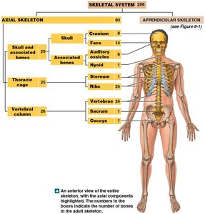
The axial skeleton forms the longitudinal axis of the human body and is essential for support, protection, and movement. It consists of 80 bones, including the skull, thoracic cage, and vertebral column.
Axial skeleton: Includes bones of the head and trunk.
Appendicular skeleton: Includes bones of the limbs and girdles.
Components of the Axial Skeleton
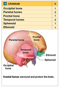
Skull: 8 cranial bones, 14 facial bones
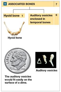
Associated bones: 6 auditory ossicles, 1 hyoid bone
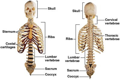
Thoracic cage: Sternum and 24 ribs
Vertebral column: 24 vertebrae, sacrum, coccyx
Functions of the Axial Skeleton
Support and protection: Brain, spinal cord, thoracic and abdominal organs
Attachment points: Muscles for head, neck, trunk movement, breathing, and stabilization of appendicular skeleton
The Skull
Cranial and Facial Bones
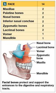
The skull is composed of 22 bones: 8 cranial bones that protect the brain and 14 facial bones that support entrances to the digestive and respiratory tracts.
Cranial bones: Occipital, parietal (2), frontal, temporal (2), sphenoid, ethmoid
Facial bones: Maxillae (2), palatine (2), nasal (2), vomer, inferior nasal conchae (2), zygomatic (2), lacrimal (2), mandible
Associated Bones
Auditory ossicles: Three tiny bones in each ear (malleus, incus, stapes) for hearing
Hyoid bone: Supports the larynx and attaches muscles of the tongue and pharynx
Major Sutures of the Skull
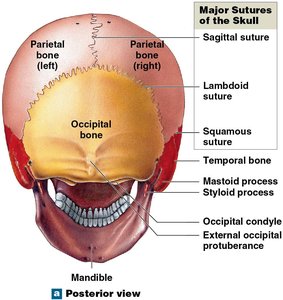
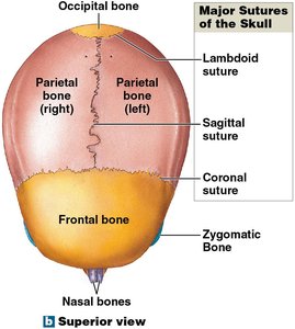
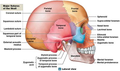
Sutures are immovable joints connecting skull bones.
Lambdoid suture: Occipital to parietal bones
Coronal suture: Frontal to parietal bones
Sagittal suture: Between parietal bones
Squamous suture: Temporal to parietal bones
Bone Markings and Foramina
Bone markings include foramina (openings for nerves and blood vessels), fissures, and sinuses.
Sinuses: Air-filled chambers that lighten bone, moisten air, and act as resonating chambers for speech
Foramina: Passageways for nerves and vessels (e.g., supra-orbital foramen, optic canal, foramen magnum)
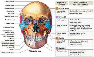
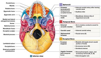
Sectional Anatomy of the Skull
Sectional views reveal the spatial relationships of cranial and facial bones, sinuses, and foramina. 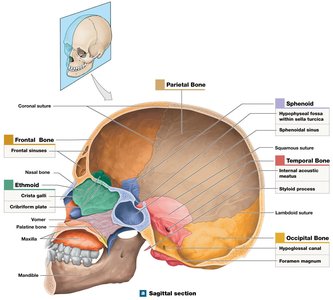
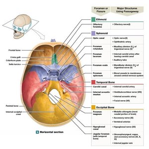
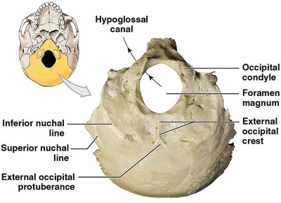
Occipital Bone
Location: Posterior and inferior surfaces of cranium
Markings: External occipital protuberance, occipital condyles, nuchal lines
Foramina: Foramen magnum, jugular foramen, hypoglossal canals
Parietal Bones
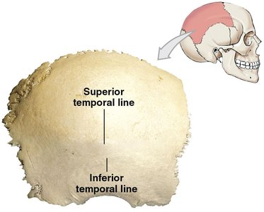
Location: Superior and lateral surfaces of cranium
Markings: Superior and inferior temporal lines, grooves for cranial blood vessels
Frontal Bone
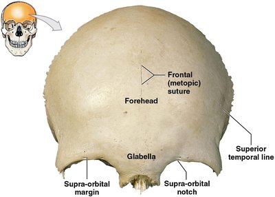
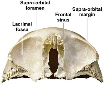
Location: Anterior cranium, roof of eye sockets
Markings: Forehead, supra-orbital margin, glabella, lacrimal fossa, frontal sinuses
Foramina: Supra-orbital foramen/notch
Facial Bones
Maxillae
Functions: Support upper teeth, form orbital rims, upper jaw, hard palate, contain maxillary sinuses
Markings: Orbital rim, anterior nasal spine, alveolar process, palatine processes, nasolacrimal canal
Foramina: Infra-orbital foramen, inferior orbital fissure
Palatine Bones
Functions: Form posterior hard palate, contribute to orbit floors
Markings: Horizontal plate, perpendicular plate
Foramina: Lateral foramina for nerves and vessels
Nasal Bones
Functions: Support bridge of nose, connect to nasal cartilage
Vomer
Function: Forms inferior portion of bony nasal septum
Inferior Nasal Conchae
Functions: Slow inhaled air, create turbulence, increase surface area for warming/humidifying air
Zygomatic Bones
Functions: Form cheekbones, rims/lateral walls of orbits, zygomatic arches
Markings: Temporal process
Foramina: Zygomaticofacial foramen
Lacrimal Bones
Function: Form medial walls of orbits
Markings: Lacrimal sulcus
Mandible
Function: Forms lower jaw
Markings: Body, alveolar part, mental protuberance, mylohyoid line, ramus, condylar process, coronoid process, mandibular notch
Foramina: Mental foramina, mandibular foramen
Hyoid Bone
Functions: Supports larynx, attaches muscles of larynx, pharynx, tongue
Markings: Body, greater horns, lesser horns
Orbital and Nasal Complexes
Orbital Complex
The orbital complex consists of seven bones forming the bony sockets for the eyes.
Bones: Frontal, maxilla, palatine, lacrimal, ethmoid, sphenoid, zygomatic
Nasal Complex and Paranasal Sinuses
Bones: Frontal, sphenoid, ethmoid, maxillae, lacrimal, inferior nasal conchae, nasal bones
Paranasal sinuses: Frontal, ethmoidal, sphenoidal, maxillary sinuses
Function: Lighten skull, moisten/clean air, resonance for speech
Fontanelles and Skull Development
Infant Skull and Fontanelles
Infant skull: Many ossification centers, bones connected by fibrous tissue
Fontanelles: Large fibrous areas allowing for brain growth and flexibility during birth
Types: Sphenoidal, mastoid, anterior, posterior fontanelles
Vertebral Column
Curves of the Vertebral Column
The vertebral column consists of 26 bones and four curves: cervical, thoracic, lumbar, and sacral.
Primary curves: Thoracic and sacral (present at birth, accommodate organs)
Secondary curves: Cervical and lumbar (develop after birth, shift weight for upright posture)
Clinical Note: Spinal Curvature Disorders
Kyphosis: Excessive posterior thoracic curvature
Lordosis: Excessive anterior lumbar curvature
Scoliosis: Lateral curvature of the spine
Vertebral Regions and Anatomy
Cervical: 7 vertebrae (smallest, most flexible)
Thoracic: 12 vertebrae (articulate with ribs)
Lumbar: 5 vertebrae (largest, support most weight)
Sacral: Sacrum (5 fused vertebrae)
Coccygeal: Coccyx (3-5 fused vertebrae)
The Thoracic Cage
Structure and Function
The thoracic cage consists of thoracic vertebrae, ribs, costal cartilages, and sternum.
Functions: Protects thoracic organs, provides muscle attachment for breathing and movement
Ribs
True ribs: First 7 pairs, attach directly to sternum
False ribs: Pairs 8-12, do not attach directly to sternum
Floating ribs: Pairs 11-12, no sternum attachment
Sternum
Parts: Manubrium, body, xiphoid process
Functions: Articulates with clavicles and ribs, provides muscle attachment
Summary Table: Axial Skeleton Components
Region | Bones | Function |
|---|---|---|
Skull | 22 (8 cranial, 14 facial) | Protects brain, supports entrances to digestive/respiratory tracts |
Associated Bones | 7 (6 auditory ossicles, 1 hyoid) | Hearing, supports larynx/tongue |
Thoracic Cage | 25 (24 ribs, 1 sternum) | Protects thoracic organs, muscle attachment |
Vertebral Column | 26 (24 vertebrae, sacrum, coccyx) | Protects spinal cord, supports body weight |
