 Indietro
Study Guide: The Brain and Cranial Nerves (Anatomy & Physiology)
Guida di studio - Note intelligenti
 Appunti personalizzati basati sui tuoi materiali, ampliati con definizioni chiave, esempi e contesto.
The Nervous System: Overview
Divisions and Structure
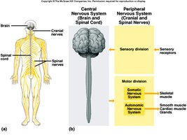
The nervous system is divided into the Central Nervous System (CNS) and Peripheral Nervous System (PNS). The CNS consists of the brain and spinal cord, while the PNS includes cranial and spinal nerves that connect the CNS to the rest of the body.
CNS: Integrates and processes information.
PNS: Transmits sensory and motor signals between the CNS and the body.
Sensory Division: Carries information from sensory receptors to the CNS.
Motor Division: Sends commands from the CNS to muscles and glands; includes somatic (voluntary) and autonomic (involuntary) systems.
Embryonic Development of the Brain
Brain Vesicles and Adult Structures
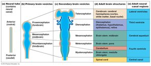
The brain develops from the neural tube, which forms primary and secondary brain vesicles. These vesicles give rise to the major regions of the adult brain.
Primary Vesicles: Prosencephalon (forebrain), Mesencephalon (midbrain), Rhombencephalon (hindbrain).
Secondary Vesicles: Telencephalon, Diencephalon, Metencephalon, Myelencephalon.
Adult Structures: Cerebrum, diencephalon, brainstem (midbrain, pons, medulla oblongata), cerebellum.
Major Regions of the Adult Brain
Structural Overview
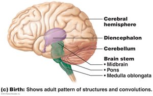
The adult brain consists of several distinct regions, each with specialized functions.
Cerebrum: Responsible for higher functions such as reasoning, memory, and voluntary movement.
Diencephalon: Includes thalamus and hypothalamus, involved in sensory relay and homeostasis.
Brain Stem: Controls basic life functions; includes midbrain, pons, and medulla oblongata.
Cerebellum: Coordinates movement and balance.
Protection of the Brain
Physical and Biochemical Barriers
The brain is protected by several mechanisms:
Skull Bones: Provide mechanical protection.
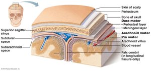
Meninges: Three connective tissue layers (dura mater, arachnoid mater, pia mater) surround the brain and spinal cord.
Cerebrospinal Fluid (CSF): Cushions and nourishes the brain.
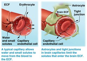
Blood-Brain Barrier: Limits passage of substances from blood to brain tissue, maintained by tight junctions and astrocytes.
Meninges of the Brain
Layers and Spaces
The meninges are three protective membranes:
Dura Mater: Tough, outer layer; forms dural folds for anchoring.
Arachnoid Mater: Web-like middle layer; contains subarachnoid space filled with CSF.
Pia Mater: Delicate inner layer; adheres to brain surface.
Subdural Space: Between dura and arachnoid; contains interstitial fluid.
Subarachnoid Space: Between arachnoid and pia; contains CSF.
Dural Folds
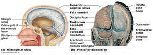
Dural folds are extensions of the dura mater that help stabilize the brain.
Falx Cerebri: Separates cerebral hemispheres.
Tentorium Cerebelli: Separates cerebellum from occipital lobes.
Ventricles of the Brain
Structure and Function
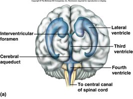
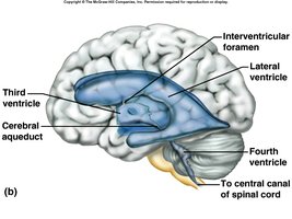
The brain contains four interconnected ventricles filled with CSF.
Lateral Ventricles (1 & 2): Located in cerebral hemispheres.
Third Ventricle: Located in diencephalon.
Fourth Ventricle: Located between brainstem and cerebellum.
Cerebral Aqueduct: Connects third and fourth ventricles.
Cerebrospinal Fluid (CSF)
Formation and Circulation
CSF is produced by the choroid plexus in the ventricles and circulates through the CNS.
Production: Choroid plexus secretes CSF (~500 ml/day).
Circulation: Flows through ventricles, central canal, and subarachnoid space.
Functions: Provides nutrition, removes waste, cushions CNS, maintains ion balance.
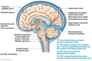
Cerebrum: Structure and Functional Areas
Lobes and Functions
The cerebrum is the largest part of the brain, divided into lobes named for overlying bones.
Frontal Lobe: Motor functions, reasoning, personality.
Parietal Lobe: Sensory processing.
Occipital Lobe: Visual processing.
Temporal Lobe: Auditory processing, memory.
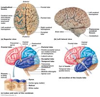
Functional Areas
The cerebrum contains motor, sensory, and association areas.
Motor Areas: Control voluntary movements (frontal cortex).
Sensory Areas: Receive and interpret sensory information (parietal, occipital, temporal cortex).
Association Areas: Integrate information for reasoning, memory, and learning.
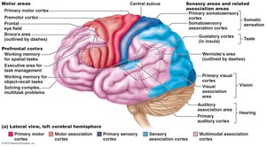
Motor/Sensory Homunculi
Homunculi are maps showing the amount of cortical tissue devoted to each body part for motor and sensory functions.
Motor Homunculus: Precentral gyrus.
Sensory Homunculus: Postcentral gyrus.
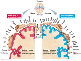
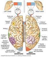
Hemispheric Lateralization
The two cerebral hemispheres are specialized for different functions.
Left Hemisphere: Language, logic, analytical tasks.
Right Hemisphere: Spatial abilities, creativity, recognition of patterns.
Basal Nuclei
Structure and Function
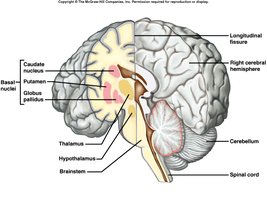
Basal nuclei are deep masses of gray matter involved in motor control.
Components: Caudate nucleus, putamen, globus pallidus.
Function: Subconscious control of learned movement patterns; relay motor impulses via thalamus.
Cerebral White Matter
Types of Tracts
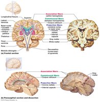
Cerebral white matter consists of myelinated tracts that connect different brain regions.
Association Tracts: Connect areas within the same hemisphere.
Commissural Tracts: Connect corresponding gyri in opposite hemispheres (e.g., corpus callosum).
Projection Tracts: Ascending and descending tracts connecting cortex with lower CNS regions.
Limbic System
Structure and Function
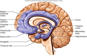
The limbic system is a group of structures involved in emotion, motivation, and memory.
Components: Portions of frontal and temporal lobes, hypothalamus, thalamus, basal nuclei, mammillary bodies, amygdala, hippocampus.
Functions: Controls emotions, produces feelings, interprets sensory impulses, facilitates memory storage and retrieval.
Diencephalon
Thalamus
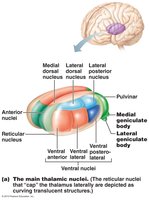
The thalamus is a major relay station for sensory information.
Functions: Relays vision, hearing, taste, pain, touch, pressure, temperature; gateway to cerebral cortex.
Structure: Forms wall of third ventricle; contains multiple nuclei.
Hypothalamus
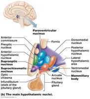
The hypothalamus regulates homeostasis and links the nervous and endocrine systems.
Functions: Controls heart rate, blood pressure, body temperature, water balance, feeding/satiety, sleep, emotional responses.
Structure: Contains multiple nuclei; connects to pituitary gland.
Brain Stem
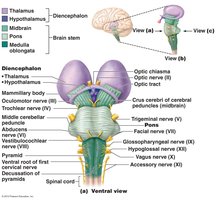
Midbrain
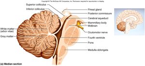
The midbrain connects the diencephalon to the pons and contains important nuclei.
Functions: Visual and auditory reflex centers; contains red nucleus and substantia nigra.
Structure: Cerebral aqueduct, cerebral peduncles, corpora quadrigemina.
Pons
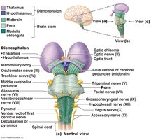
The pons is a bulge on the underside of the brainstem.
Functions: Regulates breathing; relays impulses between medulla, brainstem, and cerebellum.
Structure: Contains longitudinal and transverse tracts.
Medulla Oblongata
The medulla oblongata is the continuation of the spinal cord within the skull.
Functions: Conducts ascending and descending impulses; contains cardiac, vasomotor, and respiratory centers.
Structure: Contains nuclei for cranial nerves IX-XII.
Reticular Formation
Structure and Function
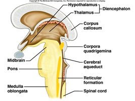
The reticular formation is a network of nerve fibers in the brainstem.
Functions: Filters sensory information, modulates pain, arouses cerebral cortex (reticular activating system).
Connections: Extends into diencephalon; connects to hypothalamus, basal nuclei, cerebellum, cerebrum.
Cerebellum
Structure and Function
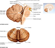
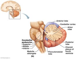
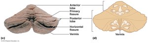
The cerebellum coordinates movement and maintains posture.
Functions: Integrates sensory information, coordinates skeletal muscle activity, helps maintain posture.
Structure: Contains anterior and posterior lobes, vermis, arbor vitae.
Cranial Nerves
Overview and Classification
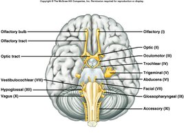
Cranial nerves are paired nerves arising from the brain, numbered I-XII.
Classification: Sensory, motor, or mixed.
Function: Innervate head, neck, and some thoracic/abdominal structures.
Trigeminal Nerve (V)
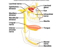
The trigeminal nerve is the major sensory nerve of the face, with three divisions.
Ophthalmic: Sensory from eyes, scalp, forehead.
Maxillary: Sensory from upper teeth, gum, lip, palate, skin of face.
Mandibular: Sensory from jaw, lower teeth, gum, lip; motor to muscles of mastication.
Facial Nerve (VII)
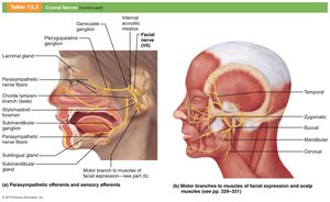
The facial nerve is the major motor nerve of the face.
Sensory: Taste from anterior 2/3 of tongue.
Motor: Muscles of facial expression, tear glands, salivary glands.
Vestibulocochlear (VIII) and Glossopharyngeal (IX)
Vestibulocochlear: Sensory from equilibrium and hearing receptors.
Glossopharyngeal: Sensory from pharynx, tonsils, tongue, carotid arteries; motor to parotid gland and pharynx muscles.
Vagus Nerve (X)
The vagus nerve innervates thoracic and abdominal viscera.
Somatic Motor: Muscles of speech and swallowing.
Autonomic Motor: Parasympathetic control of heart, lungs, digestive organs.
Sensory: Pharynx, larynx, esophagus, viscera.
Accessory (XI) and Hypoglossal (XII)
Accessory: Motor to neck and back muscles.
Hypoglossal: Motor to tongue muscles; important for speech and swallowing.
Summary Table: Cranial Nerves
Nerve | Type | Main Function |
|---|---|---|
Olfactory (I) | Sensory | Smell |
Optic (II) | Sensory | Vision |
Oculomotor (III) | Motor | Eye movement, pupil constriction |
Trochlear (IV) | Motor | Eye movement (superior oblique) |
Trigeminal (V) | Mixed | Sensory face, motor mastication |
Abducens (VI) | Motor | Eye movement (lateral rectus) |
Facial (VII) | Mixed | Facial expression, taste |
Vestibulocochlear (VIII) | Sensory | Hearing, balance |
Glossopharyngeal (IX) | Mixed | Taste, swallowing |
Vagus (X) | Mixed | Viscera, speech, swallowing |
Accessory (XI) | Motor | Neck, back muscles |
Hypoglossal (XII) | Motor | Tongue movement |
Key Review Points
The brain is protected by skull, meninges, CSF, and blood-brain barrier.
Major brain regions: cerebrum, diencephalon, brainstem, cerebellum.
Cranial nerves are classified as sensory, motor, or mixed, and innervate specific regions.
