 Indietro
Study Guide: The Cardiovascular System – Blood Vessels
Guida di studio - Note intelligenti
 Appunti personalizzati basati sui tuoi materiali, ampliati con definizioni chiave, esempi e contesto.
The Cardiovascular System: Blood Vessels
Overview of Arteries and Veins
Blood vessels are essential components of the cardiovascular system, responsible for transporting blood throughout the body, regulating blood flow, controlling blood pressure, and secreting chemicals. The vasculature is organized into two circuits: the pulmonary circuit (heart to lungs) and the systemic circuit (heart to body).
Arteries: Distribution system; carry blood away from the heart and branch into smaller vessels.
Capillaries: Exchange system; form networks for rapid exchange of gases, nutrients, and wastes.
Veins: Collection system; drain blood from capillary beds and return it to the heart.
Structure and Function of Arteries and Veins
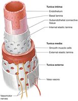
All blood vessels have a central lumen surrounded by three tissue layers called tunics:
Tunica intima: Innermost layer; composed of endothelium, subendothelial connective tissue, and internal elastic lamina. Provides a smooth surface for blood flow and elasticity for vessel stretching and recoil.
Tunica media: Middle layer; contains smooth muscle cells and external elastic lamina. Controls vessel diameter and blood flow via vasoconstriction and vasodilation, regulated by the sympathetic nervous system.
Tunica externa (adventitia): Outermost layer; dense irregular collagenous connective tissue for support and prevention of overstretching.
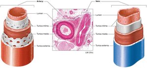
Arteries have thicker tunica media and more elastic fibers than veins, reflecting their role in blood pressure regulation and exposure to higher pressure.
Classification of Arteries and Veins
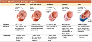
Arteries and veins are classified by size and function:
Type | Diameter | Structure | Function |
|---|---|---|---|
Elastic arteries | 2.5–1 cm | Large elastic laminae | Conduct blood under high pressure |
Muscular arteries | 1 cm–0.3 mm | Thick-walled, smooth muscle | Regulate blood flow to organs |
Arterioles | 0.3 mm–10 μm | Thin walls, few muscle cells | Control blood flow to capillary beds |
Venules | 8–100 μm | Thin walls, little muscle | Drain capillary beds |
Veins | 100 μm–1.5 cm | Thin-walled, large lumen | Return blood to heart |
Atherosclerosis
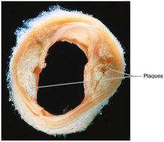
Atherosclerosis is a disease characterized by the formation of plaques (lipids, cholesterol, calcium, and debris) within the arterial tunica intima, leading to vessel narrowing and increased risk of cardiovascular events.
Vascular Anastomoses
Vascular anastomoses are connections between blood vessels, providing alternate pathways for blood flow. Arterial anastomoses are found in organs like the heart and brain, while venous anastomoses are common and form web-like patterns.
Physiology of Blood Flow
Factors That Determine Blood Pressure
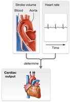
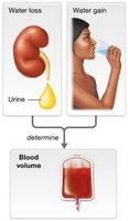
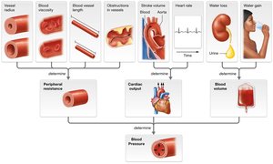
Blood pressure is influenced by three main factors: peripheral resistance, cardiac output, and blood volume.
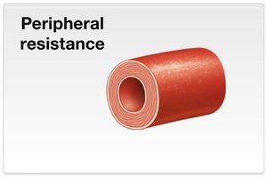
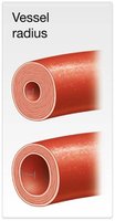
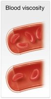
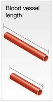
Peripheral resistance: Resistance to blood flow, greatest in the body's periphery. Increases with decreased vessel radius, increased viscosity, and increased vessel length.
Cardiac output (CO): Product of stroke volume and heart rate. $CO = SV \times HR$
Blood volume: Directly linked to water content; more volume increases pressure.
Summary Table: Factors Affecting Blood Pressure
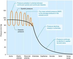
Blood Pressure in Different Portions of the Circulation
Blood pressure is low in the pulmonary circuit (~15 mm Hg) and higher in the systemic circuit (~95 mm Hg). It is highest in the aorta and elastic arteries, declining through the arterial system and veins.
Systolic pressure: ~120 mm Hg
Diastolic pressure: ~80 mm Hg
Pulse pressure: Difference between systolic and diastolic (~40 mm Hg)
Mean arterial pressure (MAP): $MAP \approx \text{Diastolic Pressure} + \frac{1}{3} \times \text{Pulse Pressure}$
Venous Return Mechanisms
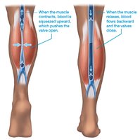
Venous valves: Prevent backward flow.
Skeletal muscle pump: Muscles contract and squeeze veins, propelling blood toward the heart.
Respiratory pump: Rhythmic changes in thoracic pressure during breathing help move blood through veins.
Capillaries and Tissue Perfusion
Capillary Structure and Function
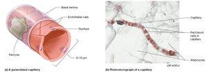
Capillaries are thin-walled vessels composed of endothelium and basal lamina. Pericytes help regulate blood flow. Capillaries are joined by tight junctions and curl around their circumference.
Types of Capillaries
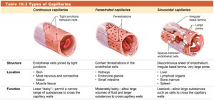
Type | Structure | Location | Function |
|---|---|---|---|
Continuous | Tight junctions | Muscle, skin, nervous tissue | Least leaky; restricts passage |
Fenestrated | Fenestrations (pores) | Kidneys, endocrine glands | Moderately leaky; rapid exchange |
Sinusoidal | Large pores, discontinuous endothelium | Liver, spleen, bone marrow | Leakiest; allows large substances |
Blood Flow through Capillary Beds
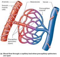
Capillary beds consist of true capillaries and a central vessel. Precapillary sphincters regulate blood flow. Thoroughfare channels drain capillary beds, and blood flows in one direction due to pressure gradients.
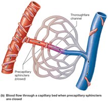
Pressures at Work in a Capillary
Water movement across capillaries is driven by hydrostatic and osmotic pressures:
Hydrostatic pressure: Force exerted by fluid; drives water out of capillary.
Osmotic pressure: Force needed to prevent water movement by osmosis; draws water into capillary, mainly due to plasma proteins (albumin).
Filtration: Passive process moving fluid from high to low pressure.
Anatomy of Systemic Arteries
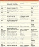
Major Systemic Arteries
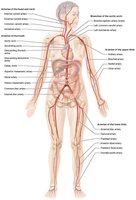
The aorta is the largest artery, with four divisions: ascending aorta, aortic arch, descending thoracic aorta, and descending abdominal aorta. Major branches supply the head, neck, thorax, abdomen, upper and lower limbs.
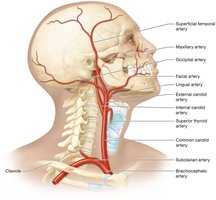
Arteries of the Head and Neck
Right and left common carotid arteries supply the head and neck, splitting into external and internal carotid arteries. External carotid branches supply superficial structures; internal carotid supplies the brain.
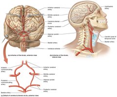
Arteries of the Thorax
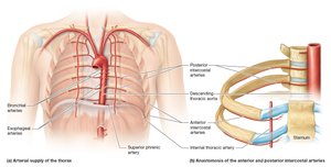
Blood supply to the thorax comes from subclavian arteries and descending thoracic aorta. Branches supply the thoracic wall, lungs, esophagus, and diaphragm.
Arteries of the Abdominal Organs
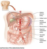
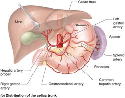
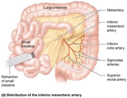
Major branches of the abdominal aorta include the celiac trunk, superior and inferior mesenteric arteries, renal arteries, and gonadal arteries. These supply the abdominal organs and viscera.
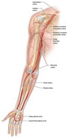
Arteries of the Upper Limb
The subclavian artery becomes the axillary and then brachial artery, supplying the upper limb. Radial and ulnar arteries terminate in palmar arches.
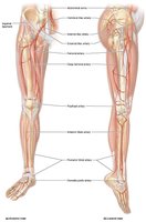
Arteries of the Lower Limb
The femoral artery is the main artery of the lower limb, becoming the popliteal artery and then splitting into anterior and posterior tibial arteries.
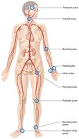
Pulse Points
Pulse points are superficial arteries where the pulse can be palpated, corresponding to heart rate and blood flow.
Major Systemic Arteries Table
Anatomy of Systemic Veins
Major Systemic Veins
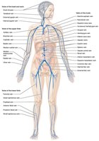
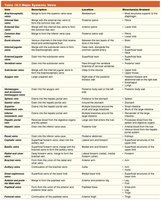
Deoxygenated blood drains into systemic veins, returning to the heart via the superior and inferior vena cava. Veins are organized by region and function.
Veins of the Head and Neck
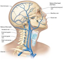
Internal and external jugular veins, along with vertebral veins, drain the head and neck, merging into the brachiocephalic vein.
Veins of the Thorax and Abdomen
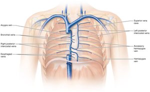
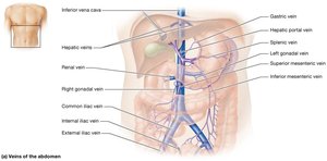
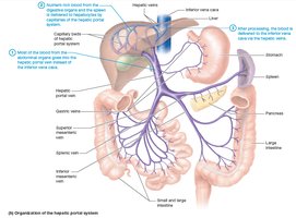
Venous drainage is complex, with the azygos system draining the posterior thoracic and abdominal walls. Pelvic organs drain into the internal iliac vein, and abdominal organs drain into the hepatic portal system for processing in the liver.
Veins of the Upper Limb
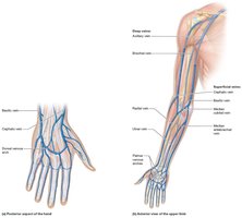
Superficial and deep venous systems drain the upper limb, with cephalic, basilic, and median veins connecting to the axillary and subclavian veins.
Veins of the Lower Limb
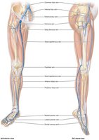
Superficial veins (great and small saphenous) and deep veins (anterior and posterior tibial, popliteal, femoral) drain the lower limb, connecting to the external iliac vein.
Major Systemic Veins Table
Vein Grafting
Vein grafting is a surgical procedure to bypass blocked arteries, often using the great saphenous vein. The vein is harvested and sewn into place to restore blood flow.
Summary
This study guide covers the anatomy and physiology of blood vessels, including their structure, function, classification, and the mechanisms regulating blood flow and pressure. It also details the major arteries and veins of the body, their regional organization, and clinical applications such as vein grafting.
