 Indietro
Study Guide: The Cardiovascular System – Blood Vessels (Anatomy & Physiology)
Guida di studio - Note intelligenti
 Appunti personalizzati basati sui tuoi materiali, ampliati con definizioni chiave, esempi e contesto.
The Cardiovascular System: Blood Vessels
Overview of Blood Vessels and Vasculature
The vasculature is a complex network of blood vessels responsible for transporting blood throughout the body. Blood vessels facilitate the exchange of gases, nutrients, and wastes, regulate blood flow, control blood pressure, and secrete various chemicals.
Arteries: Carry blood away from the heart; systemic arteries carry oxygenated blood, pulmonary arteries carry deoxygenated blood.
Capillaries: Smallest vessels; site of exchange between blood and tissues.
Veins: Return blood to the heart; systemic veins carry deoxygenated blood, pulmonary veins carry oxygenated blood.
Structure and Function of Arteries and Veins
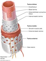
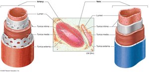
Large blood vessels are composed of three main layers, or tunics, surrounding a central lumen. Each tunic has distinct structural and functional properties.
Tunica Intima: Innermost layer; consists of endothelium (simple squamous epithelium) and basal lamina. Provides a smooth, frictionless surface and produces nitric oxide, collagen, and clotting factors.
Internal Elastic Lamina: Elastic fibers for distensibility and elasticity.
Tunica Media: Middle layer; composed of smooth muscle cells and external elastic lamina. Controls vessel diameter via vasoconstriction and vasodilation, regulated by vasomotor nerves.
Tunica Externa (Adventitia): Outermost layer; dense irregular connective tissue for support and prevention of overstretching. Contains vasa vasora—tiny vessels supplying the outer layers.
Types of Arteries
Elastic (Conducting) Arteries: Largest diameter; extensive elastic lamina; includes aorta and branches; under highest pressure.
Muscular (Distributing) Arteries: Intermediate diameter; well-developed tunica media; most named arteries; diameter changes with nervous/endocrine stimulation.
Arterioles: Smallest arteries; thin tunics; tunica media has few smooth muscle layers; regulate blood flow to capillary beds.
Types of Veins
Venules: Smallest veins; drain blood from capillary beds; postcapillary venules allow exchange.
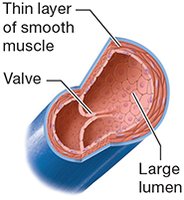
Veins: Thin walls, fewer elastic fibers, less smooth muscle; large lumens act as blood reservoirs.
Venous Valves: Extensions of tunica intima; prevent backflow; numerous in legs.
Atherosclerosis
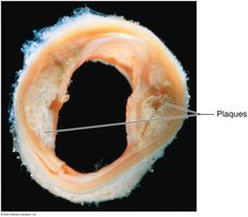
Atherosclerosis is a disease characterized by the formation of plaques within the tunica intima of arteries, leading to vessel obstruction and increased risk of heart attack or stroke.
Plaques consist of lipids, cholesterol, calcium salts, and cellular debris.
Risk factors: high blood pressure, certain cholesterol types, toxins, high blood glucose, homocysteine.
Treatment: lifestyle changes, medications, surgery.
Hemodynamics: Blood Flow and Pressure
Hemodynamics is the study of blood flow in the cardiovascular system. Blood flow is driven by a pressure gradient created by the heart and is influenced by resistance, vessel radius, viscosity, and vessel length.
Blood Pressure: Outward force exerted by blood on vessel walls; measured in mm Hg.
Blood Flow: Volume of blood per minute; directly proportional to pressure gradient, inversely proportional to resistance.
Resistance: Impedance to blood flow; increases with smaller vessel radius, higher viscosity, longer vessel length, and obstructions.
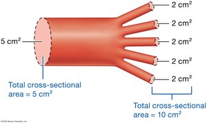
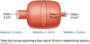
Factors Determining Blood Pressure
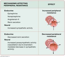
Peripheral Resistance: Most resistance occurs in the periphery; affected by vessel radius, viscosity, length, and obstructions.
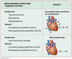
Cardiac Output (CO): Product of stroke volume and heart rate.
Blood Volume: Directly linked to water content; more water increases volume and pressure.
Compliance: Ability of vessel walls to stretch; veins are most compliant.
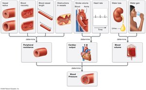
Blood Pressure in Circulation
Blood pressure varies throughout the circulatory system, being highest in the arteries and lowest in the veins.
Systolic Pressure: Pressure during ventricular systole (110–120 mm Hg).
Diastolic Pressure: Pressure during ventricular diastole (70–80 mm Hg).
Pulse Pressure: Difference between systolic and diastolic pressure (~40 mm Hg).
Mean Arterial Pressure (MAP): Average pressure during a cardiac cycle.
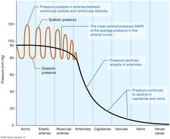
Mechanisms of Venous Return
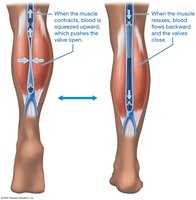
Venous Valves: Prevent backflow.
Skeletal Muscle Pump: Muscles contract and squeeze veins, propelling blood toward the heart.
Respiratory Pump: Changes in thoracic and abdominal pressure during breathing aid venous return.
Short-Term Maintenance of Blood Pressure
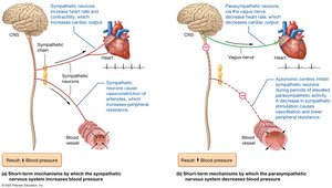
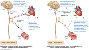
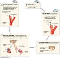
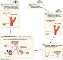
Blood pressure is regulated short-term by the nervous system and hormones, primarily by adjusting resistance and cardiac output.
Sympathetic Nervous System: Releases norepinephrine and epinephrine to increase cardiac output and vasoconstrict vessels.
Parasympathetic Nervous System: Releases acetylcholine to decrease cardiac output and induce vasodilation.
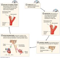
Baroreceptor Reflex: Mechanoreceptors in carotid and aortic sinuses monitor blood pressure and initiate feedback loops.
Long-Term Maintenance of Blood Pressure
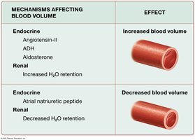
Long-term regulation is achieved by the urinary and endocrine systems, which control blood volume by adjusting water and sodium retention.
Atrial Natriuretic Peptide (ANP): Promotes excretion of water and sodium, lowering blood volume and pressure.
Antidiuretic Hormone (ADH): Increases water retention, raising blood volume and pressure.
Renin-Angiotensin-Aldosterone System (RAAS): Increases sodium and water retention, raising blood volume and pressure.
Disorders of Blood Pressure
Hypertension: Chronic high blood pressure; risk factors include genetics, age, diet, and abnormal RAAS activity.
Hypotension: Chronic or acute low blood pressure; may cause dizziness, organ failure, or shock.
Shock: Severe hypotension leading to inadequate tissue perfusion.
Capillary Structure and Function
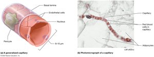
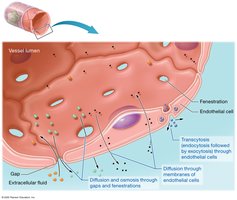
Capillaries are the site of tissue perfusion and exchange of nutrients, gases, and wastes. Their walls are thin, consisting of endothelium and basal lamina, allowing efficient exchange.
Pericytes: Contractile fibers around capillaries that regulate blood flow.
Capillary Exchange: Occurs via diffusion, osmosis, and transcytosis.
Types of Capillaries
Continuous Capillaries: Least leaky; found in muscles, skin, nervous tissue.
Fenestrated Capillaries: Leakier; found in endocrine glands, intestines, kidneys.
Sinusoidal Capillaries: Most leaky; found in liver, spleen, bone marrow.
Blood Flow through Capillary Beds
Microcirculation: Flow through capillary beds; includes true capillaries and central vessels.
Precapillary Sphincters: Regulate blood flow into capillary beds.
Autoregulation: Myogenic and metabolic mechanisms adjust perfusion based on tissue needs.
Pressures at Work in a Capillary
Hydrostatic Pressure (HP): Drives water out of capillaries; equivalent to blood pressure.
Osmotic Pressure (OP): Draws water into capillaries; determined by solute concentration, especially proteins like albumin.
Colloid Osmotic Pressure (COP): Difference in osmotic pressure between blood and interstitial fluid.
Net Filtration Pressure (NFP): Overall pressure driving water movement; calculated as
Edema
Edema: Excess water in interstitial fluid; caused by increased hydrostatic pressure or decreased colloid osmotic pressure.
Peripheral Edema: Common in lower limbs due to gravity and hypertension.
Ascites: Fluid accumulation in abdomen, often due to liver failure or malnutrition.
Anatomy of Systemic Arteries and Veins
The systemic arteries and veins are organized to supply and drain all regions of the body. The aorta is the largest artery, branching into major vessels for the head, neck, thorax, abdomen, and limbs. Veins mirror arteries, returning blood to the heart via the superior and inferior vena cava.
Aorta: Ascending, arch, descending thoracic, and abdominal segments.
Major branches: Brachiocephalic, carotid, subclavian, iliac, femoral, radial, ulnar, tibial arteries.
Veins: Jugular, subclavian, iliac, femoral, saphenous, brachial, cephalic, basilic veins.
Hepatic Portal System: Specialized circuit for detoxification and nutrient processing in the liver.
Pulse Points
Pulse: Pressure changes in arteries; palpated at superficial arteries.
Common pulse points: Carotid, radial, brachial, femoral, dorsalis pedis, posterior tibial.
Vein Grafting
Bypass Graft: Used to restore blood flow when arteries are blocked; great saphenous vein commonly used.
The Big Picture of Blood Vessel Anatomy
Blood vessels are essential for maintaining homeostasis, delivering nutrients, removing wastes, and regulating blood pressure. Their structure and function are tightly regulated by neural, endocrine, and local mechanisms to ensure proper tissue perfusion and overall cardiovascular health.
Type of Vessel | Main Function | Key Structural Features |
|---|---|---|
Artery | Carry blood away from heart | Thick tunica media, elastic laminae |
Vein | Return blood to heart | Thin walls, large lumen, valves |
Capillary | Exchange substances | Thin endothelium, pores/fenestrations |
Additional info: This guide expands on brief points with academic context, definitions, and examples to ensure completeness and clarity for exam preparation.
