 Indietro
Study Guide: The Cardiovascular System – The Heart (Anatomy & Physiology)
Guida di studio - Note intelligenti
 Appunti personalizzati basati sui tuoi materiali, ampliati con definizioni chiave, esempi e contesto.
The Cardiovascular System: The Heart
Overview and Learning Objectives
The heart is a muscular organ central to the cardiovascular system, responsible for pumping blood throughout the body. This study guide covers the anatomy, physiology, and electrical activity of the heart, as well as the cardiac cycle and regulation of cardiac output.
Describe the structure and location of the heart
Identify the chambers and valves
Explain the cardiac cycle
Trace the pathway of blood flow through the heart and major vessels
Describe factors that control heart rate and cardiac output
Calculate cardiac output based on heart rate and stroke volume
List the membranes and layers of the heart wall
Explain the conduction system
Understand electrocardiography and the origin of heart sounds
Heart Anatomy
Structure and Location
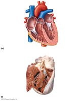
The heart is located in the mediastinum of the thoracic cavity, slightly left of the midline. It is composed of four chambers: two atria (upper chambers) and two ventricles (lower chambers). The heart is surrounded by a double-layered membrane called the pericardium.
Right Atrium: Receives deoxygenated blood from the body via the superior and inferior vena cava.
Right Ventricle: Pumps blood to the lungs via the pulmonary trunk.
Left Atrium: Receives oxygenated blood from the lungs via the pulmonary veins.
Left Ventricle: Pumps oxygenated blood to the body via the aorta.
Chambers and Valves
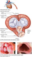
The heart contains four main valves that ensure unidirectional blood flow:
Atrioventricular (AV) Valves: Tricuspid (right) and Mitral/Bicuspid (left) valves separate the atria from the ventricles.
Semilunar (SL) Valves: Pulmonary (right) and Aortic (left) valves separate the ventricles from the major arteries.
The Cardiac Cycle
Phases of the Cardiac Cycle
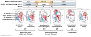
The cardiac cycle consists of all events associated with blood flow through the heart during one complete heartbeat. It includes alternating periods of contraction (systole) and relaxation (diastole).
Ventricular Filling: Blood flows passively from atria to ventricles; AV valves open, SL valves closed.
Atrial Systole: Atria contract, pushing remaining blood into ventricles.
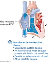
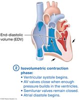
Isovolumetric Contraction: Ventricles contract, all valves closed; pressure rises, AV valves close (first heart sound: "lub").
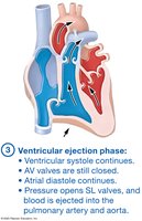
Ventricular Ejection: Ventricular pressure exceeds arterial pressure, SL valves open, blood is ejected.
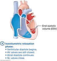
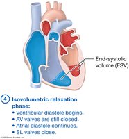
Isovolumetric Relaxation: Ventricles relax, all valves closed; SL valves close (second heart sound: "dub").
Events of the Cardiac Cycle
Pressure gradients drive unidirectional blood flow. The cycle repeats as atrial pressure exceeds ventricular pressure, opening AV valves.
Cardiac Output and Regulation
Definitions and Formulas
Cardiac Output (CO) is the amount of blood pumped by each ventricle in one minute. It is determined by heart rate (HR) and stroke volume (SV).
Stroke Volume (SV): Amount of blood pumped in one heartbeat.
Heart Rate (HR): Number of heartbeats per minute.
Cardiac Output (CO):
Example Calculation:
End-diastolic volume (EDV) = 120 mL
End-systolic volume (ESV) = 50 mL
Heart rate = 72 beats/min
Stroke volume:
Cardiac output:
Factors Influencing Stroke Volume
Preload: Degree of stretch of the ventricle (EDV).
Contractility: Strength of contraction.
Afterload: Pressure the heart must overcome to eject blood.
Ejection Fraction: (Normal: 50–65%)
Factors Influencing Heart Rate
Increased by: Sympathetic nervous system, thyroid hormone, epinephrine, norepinephrine, increased body temperature.
Decreased by: Parasympathetic nervous system, decreased body temperature.
Age & Fitness: Infants and elderly have higher HR; trained athletes have lower resting HR.
Cardiac Electrophysiology
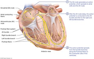
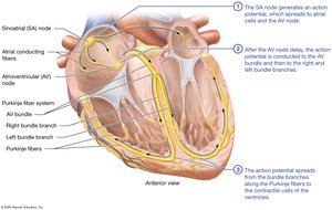
Pacemaker Cells and the Cardiac Conduction System
The heart's rhythm is set by specialized pacemaker cells, which generate and conduct electrical impulses. The conduction system ensures coordinated contraction of the heart chambers.
SA Node: Primary pacemaker, located in right atrium (60–100 bpm).
AV Node: Delays signal, located at junction of atria and ventricles (40–60 bpm).
AV Bundle, Bundle Branches, Purkinje Fibers: Conduct impulses through ventricles (25–45 bpm).
Pathway of Electrical Flow: SA node → AV node (delay) → AV bundle → Bundle branches → Purkinje fibers → Ventricles
Pacemaker Cell Action Potential
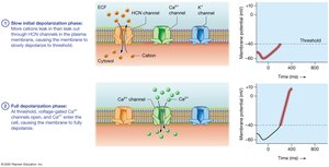
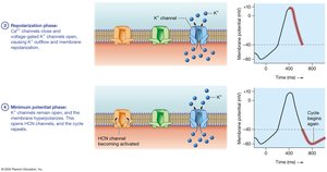
Pacemaker cells exhibit spontaneous depolarization, leading to rhythmic action potentials. Key phases include:
Initial Depolarization: Influx of cations through HCN channels.
Full Depolarization: Ca2+ influx through voltage-gated channels.
Repolarization: K+ efflux.
Minimum Potential: Cycle repeats.
Contractile Cell Action Potential
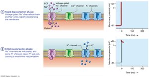
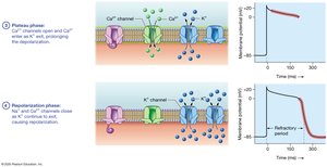
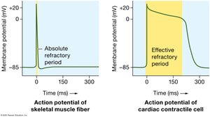
Contractile cells receive impulses from pacemaker cells and generate force. Their action potentials include a plateau phase, which distinguishes them from skeletal muscle fibers.
Rapid Depolarization: Na+ influx.
Initial Repolarization: K+ efflux.
Plateau Phase: Ca2+ influx and K+ efflux.
Repolarization: Na+ and Ca2+ channels close, K+ continues to exit.
Coronary Circulation
Arterial Supply and Venous Return
The heart muscle (myocardium) has its own blood supply and drainage system, known as the coronary circulation.
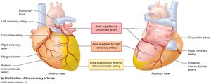
Right Coronary Artery (RCA): Supplies right atrium and ventricle.
Left Coronary Artery (LCA): Branches into LAD (anterior interventricular artery) and circumflex artery.
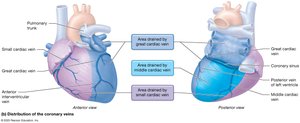
Cardiac Veins: Great, middle, and small cardiac veins drain different regions.
Coronary Sinus: Collects venous blood and empties into right atrium.
Flow: Ascending aorta → coronary arteries → myocardial capillaries → cardiac veins → coronary sinus → right atrium
Electrocardiography (ECG)
ECG Waves and Intervals
An electrocardiogram (ECG) records the electrical activity of the heart. Key components include:
P Wave: Atrial depolarization.
QRS Complex: Ventricular depolarization (and atrial repolarization, obscured).
T Wave: Ventricular repolarization.
Intervals and Segments:
R-R Interval: Duration of action potential spread; determines heart rate.
P-R Interval: Time for depolarization from SA node to ventricles; includes AV node delay.
Q-T Interval: Ventricular cells undergoing action potentials.
S-T Segment: Plateau phase; clinical changes indicate pathology.
Dysrhythmia: Disturbance in the electrical rhythm of the heart.

Cardiac Dysrhythmias
Types of Dysrhythmias
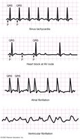
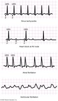
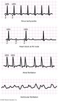
Cardiac dysrhythmias are abnormal patterns of electrical activity in the heart. They are classified as:
Disturbances in Heart Rate: Bradycardia (HR < 60 bpm), Tachycardia (HR > 100 bpm).
Disturbances in Conduction Pathways: Heart Block (blockages, often at AV node, longer P-R interval, extra P waves).
Fibrillation: Atrial Fibrillation (irregular rhythm, not life-threatening), Ventricular Fibrillation (life-threatening, treated with defibrillation), Asystole (no electrical activity).



Summary Table: Cardiac Cycle Phases
Phase | Chamber Activity | AV Valves | SL Valves | Key Event |
|---|---|---|---|---|
Ventricular Filling | Atria & ventricles relaxed | Open | Closed | 80% blood flows passively |
Atrial Systole | Atria contracted, ventricles relaxed | Open | Closed | Remaining 20% blood pumped |
Isovolumetric Contraction | Ventricles contracted | Closed | Closed | First heart sound (lub) |
Ventricular Ejection | Ventricles contracted | Closed | Open | Blood ejected |
Isovolumetric Relaxation | Ventricles relaxed | Closed | Closed | Second heart sound (dub) |
Additional info:
Cardiac output is a critical measure of heart function and is used clinically to assess cardiovascular health.
ECG abnormalities can indicate underlying heart disease, electrolyte imbalances, or conduction system defects.
Coronary circulation is essential for nourishing the myocardium and preventing ischemic heart disease.
