Indietro
Study Guide: The Central Nervous System (CNS)
Guida di studio - Note intelligenti
 Appunti personalizzati basati sui tuoi materiali, ampliati con definizioni chiave, esempi e contesto.
The Central Nervous System (CNS)
Introduction to the CNS
The central nervous system (CNS) is composed of the brain and spinal cord, serving as the main integration and control center of the body. It receives sensory information from the peripheral nervous system, processes and interprets this information, and coordinates motor responses that travel back through descending pathways.
Integration: The CNS processes incoming sensory data.
Motor Coordination: Initiates and sends out motor commands.
Communication: Connects with the peripheral nervous system for sensory input and motor output.
Major Divisions of the Brain
Brain Areas and Their Functions
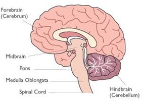
The brain is divided into four main regions, each with distinct functions:
Cerebrum: Responsible for conscious thought, reasoning, memory, sensory interpretation, and voluntary movement. It is the largest region.
Diencephalon: Contains the thalamus, hypothalamus, and epithalamus; involved in sensory relay and homeostasis.
Brainstem: Connects the brain to the spinal cord and controls essential automatic functions (midbrain, pons, medulla oblongata).
Cerebellum: Coordinates movement, posture, and balance.
The Cerebrum
Anatomy and Function
The cerebrum is the largest region, divided into right and left hemispheres. It is responsible for conscious awareness, reasoning, memory, voluntary movement, and sensory processing. The outer layer, the cerebral cortex, is composed of gray matter (neuronal cell bodies), while deeper regions contain white matter (myelinated axons).
Gray Matter: Processing and integration of information.
White Matter: Transmission of information between brain regions.
Surface Anatomy: Gyri, Sulci, and Fissures
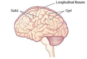
The surface of the cerebrum features:
Gyri: Raised folds or ridges.
Sulci: Shallow grooves separating gyri.
Fissures: Deep grooves separating major regions.
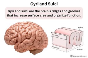
Importance of Gyri and Sulci
The folds created by gyri and sulci increase the surface area of the cerebral cortex, allowing more neurons to fit within the skull and enhancing the brain's processing capacity.
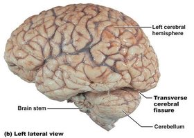
Major Fissures of the Cerebrum
Longitudinal fissure: Separates right and left hemispheres.
Transverse cerebral fissure: Separates cerebrum from cerebellum.
Sylvain fissure (lateral sulcus): Separates temporal lobe from frontal and parietal lobes.
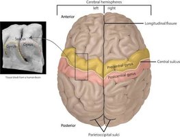
The Central Sulcus
The central sulcus separates the frontal lobe from the parietal lobe. The precentral gyrus (frontal side) is involved in motor control, while the postcentral gyrus (parietal side) is involved in sensory processing.
The Four Major Lobes of the Cerebrum
Lobe Functions
Frontal Lobe: Voluntary motor control, planning, judgment, speech production (Broca's area).
Parietal Lobe: Processes touch, pain, temperature, and body position.
Temporal Lobe: Hearing, memory, aspects of language comprehension, olfaction.
Occipital Lobe: Processes visual information.
Functional Areas of the Cerebral Cortex
Motor, Sensory, and Association Areas
Motor Area: Controls voluntary skeletal muscle movement (primarily frontal lobe).
Sensory Area: Receives and interprets sensory information (parietal, temporal, occipital lobes).
Association Areas: Integrates information, involved in reasoning, memory, language, and decision making (spread throughout cortex).
Premotor Cortex, Primary Motor Cortex, and Somatosensory Cortex
Premotor Cortex: Plans and prepares voluntary movements (frontal lobe).
Primary Motor Cortex: Initiates voluntary movements (precentral gyrus).
Supplemental Motor Cortex: Organizes and coordinates movements involving both sides of the body.
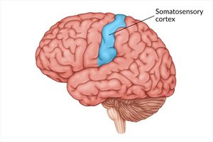
Primary Somatosensory Cortex: Receives and processes touch, pressure, pain, temperature, and body position (postcentral gyrus).
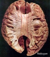
The Corpus Callosum
Structure and Function
The corpus callosum is a large bundle of white matter fibers connecting the right and left cerebral hemispheres, allowing communication and coordination of sensory, motor, and cognitive abilities.
White Matter: Composed of myelinated fibers.
Example: Coordinated use of both hands requires communication between hemispheres.
The Diencephalon
Anatomy and Functions
The diencephalon is located between the cerebral hemispheres and above the midbrain. It serves as a major sensory relay and regulatory center.
Thalamus: Major sensory relay station (except smell).
Hypothalamus: Regulates homeostasis, body temperature, hunger, thirst, autonomic activity, and hormone secretion.
Epithalamus: Contains pineal gland, produces melatonin, regulates sleep-wake cycle.
The Brainstem
Anatomy and Major Functions
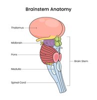
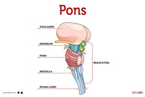
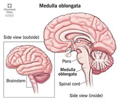
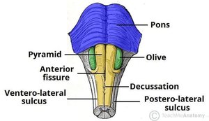
The brainstem connects the brain to the spinal cord and consists of the midbrain, pons, and medulla oblongata. It provides pathways for sensory and motor information and regulates essential involuntary functions.
Midbrain: Visual and auditory reflexes, motor control.
Pons: Communication, respiratory regulation, sleep regulation.
Medulla oblongata: Cardiovascular and respiratory regulation, protective reflexes.
Midbrain Functions
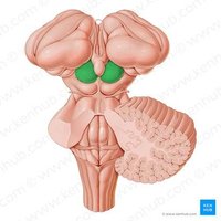
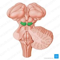
Visual Reflexes: Superior colliculi coordinate reflexive eye and head movement.
Auditory Reflexes: Inferior colliculi process sound and orient head/eyes toward source.
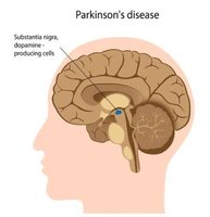
Motor Control: Substantia nigra produces dopamine for smooth, controlled movements.
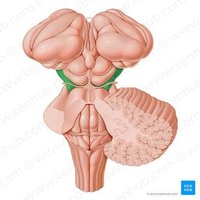
Motor Pathways: Cerebral peduncles carry descending motor fibers.
Parkinson's Disease
Parkinson's disease is a progressive neurological disorder caused by degeneration of dopamine-producing neurons in the substantia nigra. Dopamine helps the basal nuclei select appropriate movements and suppress unwanted ones.
Symptoms: Resting tremors, muscle rigidity, bradykinesia (slowness of movement), postural instability.
Pons Functions
Communication: Relays information between cerebrum, cerebellum, and CNS.
Respiratory Regulation: Regulates breathing rate and pattern.
Sleep Regulation: Participates in sleep-wake cycles, especially REM sleep.
Cranial Nerves: Nuclei for cranial nerves V-VIII.
Medulla Oblongata Functions
Cardiovascular Regulation: Controls heart rate and blood vessel diameter.
Respiratory Regulation: Generates and regulates basic breathing rhythm.
Protective Reflexes: Controls swallowing, coughing, sneezing, gagging, vomiting.
Sensory & Motor Pathways: Carries ascending sensory and descending motor commands.
Pyramidal Decussation: Most descending motor fibers cross to the opposite side.
Reticular Formation
Anatomy and Functions
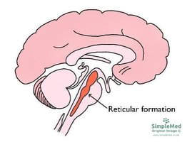
The reticular formation is a network of neurons extending throughout the brainstem. It is involved in consciousness, alertness, sensory filtering, sleep-wake cycle, motor regulation, and autonomic regulation.
Consciousness: Maintains wakefulness and awareness.
Sensory Filtering: Filters incoming sensory information.
Motor Regulation: Regulates muscle tone and posture.
Autonomic Regulation: Contributes to respiratory and cardiovascular control.
Cranial Nerves and the Brainstem
Relationship and Functions
There are 12 pairs of cranial nerves carrying sensory and/or motor information, primarily between the brain and head/neck structures. Most are associated with nuclei in the brainstem.
Midbrain: Cranial nerves III & IV
Pons: Cranial nerves V-VIII
Medulla: Cranial nerves IX-XII
Forebrain: Cranial nerves I & II
Cerebellum: Anatomy & Organization
Major Features
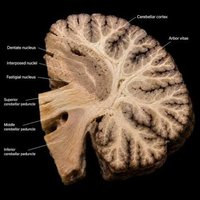
The cerebellum is located posterior to the pons and medulla, inferior to the occipital lobes. It consists of two hemispheres, vermis, cerebellar cortex (gray matter), arbor vitae (white matter), cerebellar peduncles, and deep cerebellar nuclei.
Lobes of the Cerebellum
Anterior Lobe: Regulates posture, muscle tone, trunk and limb movement.
Posterior Lobe: Plans and coordinates skilled voluntary movements.
Flocculonodular Lobe: Maintains balance, equilibrium, coordinates eye movements.
Functions of the Cerebellum
Coordination: Ensures movements are smooth and controlled.
Balance and Equilibrium: Integrates vestibular information.
Posture and Muscle Tone: Regulates muscle activity for stability.
Motor Learning: Refines movements through practice.
Error Correction: Compares intended and actual movements.
Protection of the CNS
Protective Mechanisms
Bone: Skull protects the brain; vertebral column protects the spinal cord.
Meninges: Three connective tissue membranes (dura mater, arachnoid mater, pia mater) surround and protect the CNS.
Cerebrospinal Fluid (CSF): Cushions the CNS, acts as a shock absorber, provides a stable chemical environment.
Blood-Brain Barrier: Selectively controls movement of substances from blood to brain.
Meninges of the CNS
Dura mater: Tough, outermost membrane.
Arachnoid mater: Thin, web-like middle membrane.
Pia mater: Delicate, innermost membrane.
Subarachnoid space: Contains CSF, located between arachnoid and pia mater.
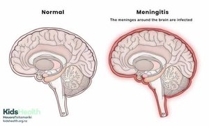
Meningitis
Definition, Types, and Symptoms
Meningitis is inflammation of the meninges. It can be bacterial (more severe, medical emergency) or viral (generally milder).
Symptoms: Severe headache, fever, stiff neck, photophobia.
Complications: Increased intracranial pressure, brain swelling, seizures, neurological damage.