 Indietro
Ch. 13 - The Central Nervous System (CNS) – Structure, Function, and Development
Guida di studio - Note intelligenti
 Appunti personalizzati basati sui tuoi materiali, ampliati con definizioni chiave, esempi e contesto.
The Central Nervous System (CNS)
Overview and Directional Terms
The central nervous system (CNS) consists of the brain and spinal cord, serving as the main control center for the body. Unique directional terms are used in CNS anatomy:
Rostral: Toward the nose (anterior)
Caudal: Toward the tail (posterior)
The Brain
Functions and Control
The brain regulates vital functions such as heart rate, respiratory rate, and blood pressure. It controls the autonomic nervous system and endocrine system, and innervates the head via cranial nerves. The brain is responsible for complex neural functions including intelligence, consciousness, memory, sensory-motor integration, emotion, behavior, and socialization.
Embryonic Development of the Brain
Primary and Secondary Brain Vesicles
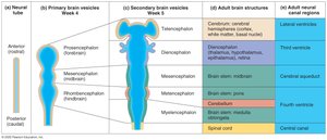
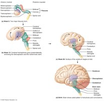
Brain development begins from the rostral part of the neural tube. In a 4-week-old embryo, three primary brain vesicles form:
Prosencephalon: Forebrain
Mesencephalon: Midbrain
Rhombencephalon: Hindbrain
These vesicles further divide into secondary vesicles, which give rise to adult brain structures:
Telencephalon: Cerebral hemispheres
Diencephalon: Thalamus, hypothalamus, epithalamus
Metencephalon: Pons and cerebellum
Myelencephalon: Medulla oblongata
Basic Parts and Organization of the Brain
Brain Regions
The brain is classified into four main regions:
Brain stem: Midbrain, pons, medulla oblongata
Cerebellum
Diencephalon
Cerebrum: Two hemispheres, four lobes
Ventricles of the Brain
Structure and Function
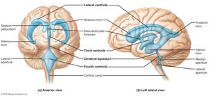
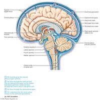
The brain contains four ventricles, which are expansions of the central cavity filled with cerebrospinal fluid (CSF) and lined with ependymal cells. They are continuous with each other and with the central canal of the spinal cord:
Lateral ventricles: Located in cerebral hemispheres, horseshoe-shaped
Third ventricle: Located in the diencephalon, connected to lateral ventricles by the interventricular foramen
Cerebral aqueduct: Connects third and fourth ventricles
Fourth ventricle: Located in the hindbrain, connects to the central canal of the spinal cord
Distribution of Gray and White Matter
Organization in the CNS
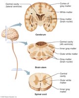
Gray matter, containing neuronal cell bodies, is centrally located, while white matter, containing axons, is external. An additional layer of gray matter, called the cortex, is found in the cerebrum and cerebellum.
The Brain Stem
Structure and Functions
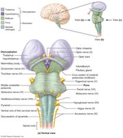
The brain stem includes the midbrain, pons, and medulla oblongata. It serves as a passageway for fiber tracts between the cerebrum and spinal cord, is involved in face and head innervation, and produces automatic behaviors necessary for survival. It integrates auditory and visual reflexes, and ten pairs of cranial nerves attach to it.
The Cerebellum
Structure and Function
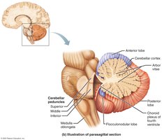
The cerebellum is located dorsal to the pons and medulla, and is responsible for smoothing and coordinating body movements and maintaining equilibrium. It consists of two hemispheres, anterior and posterior lobes, and its surface is folded into ridges called folia.
Cortex: Gray matter
Arbor vitae: Internal white matter
Deep cerebellar nuclei
The cerebellum receives information on equilibrium, current movements, and instructions from the cerebral cortex to coordinate and fine-tune motor commands. It also participates in cognition, language, and problem solving.
The Diencephalon
Structure and Components
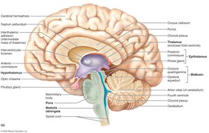
The diencephalon forms the central core of the forebrain and is composed of three paired structures:
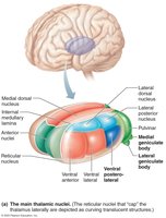
Thalamus: Relay station for sensory information
Hypothalamus: Main visceral control center
Epithalamus: Includes the pineal gland, which secretes melatonin
The Cerebrum
Lobes, Sulci, and Gyri
The cerebrum accounts for 83% of brain mass and is divided into lobes by deep grooves called fissures and sulci. The lobes are named for the overlying skull bones: frontal, parietal, occipital, and temporal. The central sulcus separates the frontal and parietal lobes, bordered by the precentral and postcentral gyri.
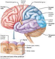
The Cerebral Cortex
Functional Regions
The cerebral cortex is the home of conscious mind, enabling awareness, voluntary movement, communication, memory, and understanding. It is composed of gray matter and constitutes about 40% of the brain's mass. Functional regions include:
Sensory areas: Receive and process sensory information
Association areas: Integrate sensory input
Motor areas: Plan and initiate voluntary movements
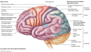
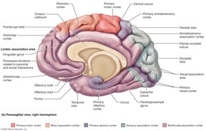
Sensory Areas
Primary Somatosensory Cortex
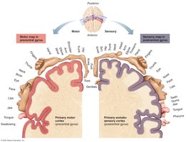
Located along the postcentral gyrus, the primary somatosensory cortex is involved in conscious awareness of general somatic senses and spatial discrimination. Projection is contralateral, meaning each hemisphere receives input from the opposite side of the body. The sensory homunculus is a body map of the sensory cortex.
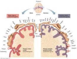
Somatosensory Association Cortex
This area lies posterior to the primary somatosensory cortex and integrates different sensory inputs, such as touch and pressure, drawing upon stored memories to recognize objects by feel.
Visual, Auditory, Vestibular, Gustatory, and Olfactory Areas
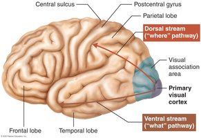
Primary visual cortex: Located in the occipital lobe, receives visual information from the retina
Visual association area: Surrounds the primary visual cortex, analyzes color, form, and movement
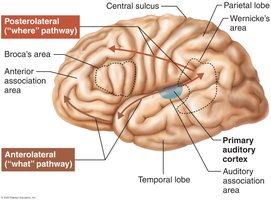
Primary auditory cortex: Located in the temporal lobe, conscious awareness of sound
Auditory association area: Permits evaluation of different sounds
Vestibular cortex: Located in the insula, conscious awareness of balance
Gustatory cortex: Located in the insula, conscious awareness of taste
Olfactory cortex: Located in the piriform cortex, conscious awareness of smells
Motor Areas
Premotor and Primary Motor Cortex
The premotor cortex, located anterior to the precentral gyrus, controls complex movements and plans voluntary actions. The primary motor cortex, located in the precentral gyrus, contains pyramidal cells that control specific areas of the body. Motor functions are contralateral, with pyramidal axons crossing to the opposite side of the brain.
Frontal Eye Field and Broca’s Area
Frontal eye field: Controls voluntary movement of the eyes
Broca’s area: Manages speech production, located in the left hemisphere
Multimodal Association Areas
Posterior, Anterior, and Limbic Association Areas
Multimodal association areas receive input from multiple sensory modalities and association areas, integrating information for unified perception, motor planning, and emotional processing.
Posterior association area: Integrates sensory information, spatial awareness, language comprehension
Anterior association area (prefrontal cortex): Involved in thinking, reasoning, judgment, impulse control, and social skills
Limbic association area: Involved with memory and emotions
Lateralization of Cortical Functioning
The two cerebral hemispheres control opposite sides of the body and are specialized for different cognitive functions. The left hemisphere is dominant for language, math, and logic, while the right hemisphere is involved in visual-spatial skills, intuition, emotion, and artistic abilities.
Cerebral White Matter
Types of Tracts
Commissural fibers: Connect the two hemispheres (e.g., corpus callosum)
Association fibers: Connect different parts of the same hemisphere
Projection fibers: Run vertically, connecting the cortex with lower regions
Deep Gray Matter of the Cerebrum
Basal Nuclei and Basal Forebrain Nuclei
Basal nuclei (ganglia) are involved in motor control, including the caudate nucleus, putamen, and globus pallidus. Basal forebrain nuclei are associated with memory, arousal, and motor control, and their degeneration is linked to Alzheimer’s disease.
Functional Brain Systems
Limbic System and Reticular Formation
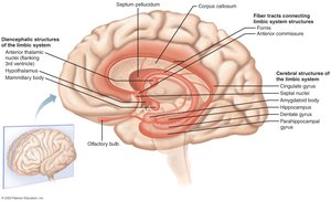
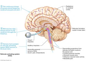
The limbic system, located in the medial aspect of the cerebral hemispheres and diencephalon, is involved in emotion and memory. The reticular formation, spanning the brain stem, maintains consciousness and alertness.
Protection of the Brain
Meninges, CSF, and Blood-Brain Barrier
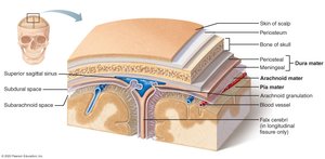
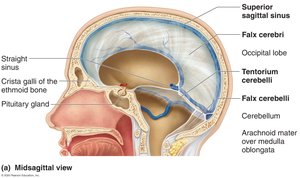
The brain is protected by the skull, meninges, cerebrospinal fluid, and the blood-brain barrier. The meninges include:
Dura mater: Strongest, two layers, encloses dural sinuses
Arachnoid mater: Beneath dura, contains villi for CSF absorption
Pia mater: Delicate, clings to brain surface
Cerebrospinal Fluid (CSF)
CSF fills the cavities of the brain and spinal cord, providing a cushion, nourishment, waste removal, and chemical signaling. It is formed in choroid plexuses in the ventricles.
Blood-Brain Barrier
The blood-brain barrier prevents most blood-borne toxins from entering the brain, but allows passage of nutrients, alcohol, nicotine, and anesthetics.
Spinal Cord
Structure and Function
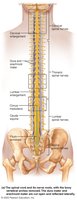
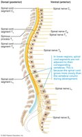
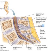
The spinal cord provides a two-way conduction pathway and is a major center for reflexes. It extends from the foramen magnum to the level of L1 or L2 vertebra. Key features include:
Conus medullaris: Inferior end
Filum terminale: Connective tissue attaching to coccyx
Cervical and lumbar enlargements: Where nerves for limbs arise
Cauda equina: Collection of spinal nerve roots
White and Gray Matter of the Spinal Cord
Organization
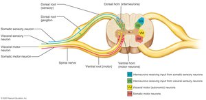
White matter is composed of myelinated and nonmyelinated axons, allowing communication between the spinal cord and brain. Fibers are classified as ascending, descending, or commissural. Gray matter is shaped like an "H" and contains interneurons and motor neuron cell bodies.
Protection of the Spinal Cord
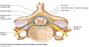
The spinal cord is protected by vertebrae, meninges, and CSF. The meninges include a single-layer dura mater, arachnoid mater, and pia mater, which extends to the coccyx and forms denticulate ligaments.
Sensory and Motor Pathways in the CNS
Ascending and Descending Pathways
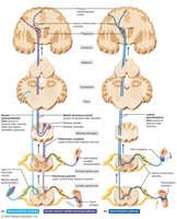
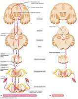
Multineuron pathways connect the brain and body periphery. Ascending pathways carry sensory information to the brain, while descending pathways deliver motor instructions from the brain to the spinal cord.
Ascending pathways: Dorsal column, spinothalamic, spinocerebellar
Descending pathways: Direct (pyramidal tracts), indirect (tectospinal, vestibulospinal, rubrospinal, reticulospinal)
Disorders of the Central Nervous System
Spinal Cord Damage and Brain Dysfunction
Paralysis: Loss of motor function
Parasthesia: Loss of sensation
Paraplegia: Injury between T1 and L2, paralysis of lower limbs
Quadriplegia: Injury in cervical region, paralysis of all four limbs
Cerebrovascular accident (stroke): Blockage or interruption of blood flow to a brain region
Alzheimer’s disease: Progressive degenerative disease leading to dementia
Congenital malformations: Neural tube defects (anencephaly, spina bifida), cerebral palsy
Additional info: The notes above expand on brief points with academic context, definitions, and examples to ensure completeness and clarity for college-level Anatomy & Physiology students.
