 Indietro
Study Guide: The Central Nervous System (CNS) – Structure, Function, and Protection
Guida di studio - Note intelligenti
 Appunti personalizzati basati sui tuoi materiali, ampliati con definizioni chiave, esempi e contesto.
Overview of the Central Nervous System
Introduction to CNS
The Central Nervous System (CNS) consists of the brain and spinal cord. It is responsible for movement, interpreting sensory information, maintaining homeostasis, and higher mental functions such as learning and language.
Motor functions: Stimulation of muscle contraction or gland secretion (performed by PNS).
Sensory functions: Detection of sensations inside and outside the body (performed by PNS).
Integrative functions: Decision-making processes, interpretation of sensory information, planning movement, maintaining homeostasis, and higher mental functions (performed by CNS).
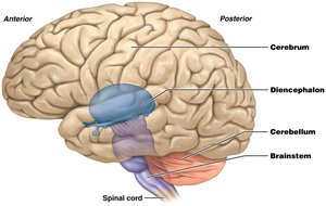
Basic Structure of the Brain and Spinal Cord
The brain is a soft, whitish-gray organ located in the cranial cavity, continuous with the spinal cord. It receives about 20% of total blood flow during rest, reflecting its high demand for oxygen and nutrients. Internal cavities called ventricles are filled with cerebrospinal fluid (CSF).
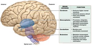
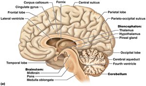
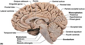
The brain consists of four main divisions: Cerebrum, Diencephalon, Cerebellum, and Brainstem.
The spinal cord is a long, tubular organ enclosed within the vertebral cavity, ending between the first and second lumbar vertebrae.
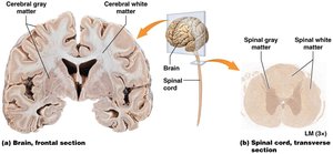
Gray Matter and White Matter
Gray matter consists of neuron cell bodies, dendrites, and unmyelinated axons. White matter consists of myelinated axons. The organization of gray and white matter is reversed in the brain and spinal cord: in the brain, white matter is deep; in the spinal cord, it is superficial.
Gray matter processes information; white matter shuttles information to and from the brain.
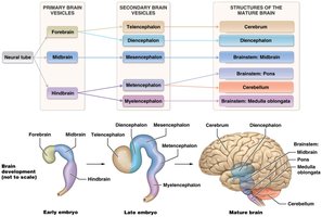
Development of the CNS
Neural Tube and Brain Vesicles
The CNS develops from a hollow structure called the neural tube. By the 4th week, the caudal end becomes the spinal cord, and the cranial end forms three primary brain vesicles: forebrain, midbrain, and hindbrain. These give rise to five secondary brain vesicles, which become the four divisions of the mature brain.
The cavity within the neural tube becomes the ventricles in the brain and the central canal in the spinal cord.
The Cerebrum
Gross Anatomy
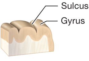
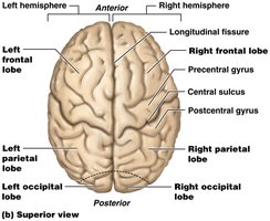
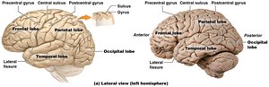
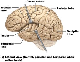
The cerebrum is the largest part of the brain, divided into left and right hemispheres. Each hemisphere is further divided into five lobes: frontal, parietal, temporal, occipital, and insula. The surface features include gyri (ridges) and sulci (grooves), which increase surface area.
Frontal lobe: Planning and executing movement, complex mental functions.
Parietal lobe: Processing and integrating sensory information, attention.
Temporal lobe: Hearing, language, memory, emotions.
Occipital lobe: Vision.
Insula: Taste and visceral functions.
Cerebral Cortex and Functional Areas
The cerebral cortex is the outer layer of gray matter, responsible for conscious processes. Most of the cortex is neocortex, composed of six layers. It contains three main functional areas:
Primary motor cortex: Plans and executes movement.
Primary sensory cortices: Receive and process sensory input.
Association areas: Integrate information; unimodal areas process one type, multimodal areas process multiple types.
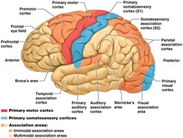
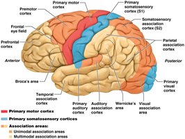
Area Name | Type of Cortex | Location | Function |
|---|---|---|---|
Primary motor cortex | Motor | Precentral gyrus of frontal lobe | Plans and executes movement |
Frontal eye fields | Association | Anterior to premotor cortex | Back-and-forth eye movements |
Somatosensory association cortex (S2) | Association | Posterior to S1 | Integrates somatic sensory stimuli |
Primary somatosensory cortex (S1) | Sensory | Postcentral gyrus of parietal lobe | Receives and processes somatic sensory stimuli |
Primary visual cortex | Sensory | Posterior occipital lobe | Processes visual stimuli |
Primary auditory cortex | Sensory | Superior temporal lobe | Processes auditory stimuli |
Gustatory cortex | Sensory | Insula and parietal lobe | Processes taste stimuli |
Vestibular areas | Sensory | Parietal and temporal lobes | Processes equilibrium and positional sense |
Olfactory cortex | Sensory | Limbic and medial temporal lobes | Processes smell stimuli |
Broca's area | Association | Anterolateral frontal lobe | Language production |
Wernicke's area | Association | Temporal and parietal lobes | Language comprehension |
Prefrontal cortex | Association | Widespread in frontal lobe | Personality, learning, higher cognition |
Parietal association cortex | Association | Widespread in parietal lobe | Integrating sensory information and attention |
Temporal association cortex | Association | Widespread in temporal lobe | Recognition and awareness |
Basal Nuclei and White Matter
Basal nuclei are clusters of neuron cell bodies deep within each cerebral hemisphere, involved in movement, behavior, cognition, and perception. The main components are the caudate nucleus, putamen, and globus pallidus.
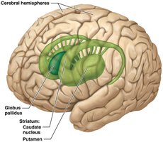
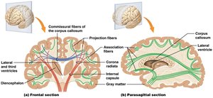
Cerebral white matter consists of three types of fibers:
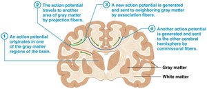
Commissural fibers: Connect right and left hemispheres (e.g., corpus callosum).
Projection fibers: Connect cortex with other brain areas and spinal cord.
Association fibers: Connect gray matter within a single hemisphere.
Limbic System
The limbic system is a functional brain system involved in memory, learning, emotion, and behavior. It includes the limbic lobe, hippocampus, amygdala, and connecting pathways.
The Diencephalon
Structure and Function
The diencephalon is located at the center of the brain and consists of four components: thalamus, hypothalamus, epithalamus, and subthalamus. Each contains nuclei that receive specific input and send output to other brain regions.
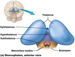
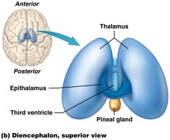
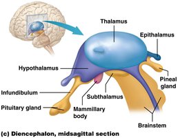
Thalamus: Main entrance to the cerebral cortex; processes and relays information.
Hypothalamus: Regulates autonomic functions, sleep/wake cycle, thirst, hunger, body temperature, and links to the pituitary gland.
Epithalamus: Contains the pineal gland, which secretes melatonin.
Subthalamus: Functionally connected with basal nuclei; controls movement.
The Cerebellum
Structure and Function
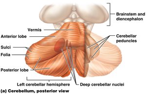
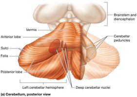
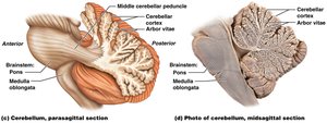
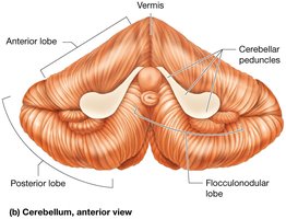
The cerebellum is located posterior and inferior to the brain. It is divided into two hemispheres connected by the vermis and three lobes: anterior, posterior, and flocculonodular. The cerebellar cortex is highly folded, and the inner white matter forms the arbor vitae.
Functions: Planning and coordination of movement, especially complex activities.
The Brainstem
Structure and Function
The brainstem connects the brain and spinal cord and is vital for immediate survival. It controls basic homeostatic functions, reflexes, movement, sensation, and alertness. The brainstem contains the midbrain, pons, and medulla oblongata.
Midbrain: Visual and auditory functions, movement regulation.
Pons: Regulation of movements, breathing, sleep, and arousal.
Medulla oblongata: Controls heart rate, breathing, blood pressure, and contains vital centers.
Protection of the Brain and Spinal Cord
Cranial Meninges
The brain is protected by three layers of meninges: dura mater, arachnoid mater, and pia mater. The dura mater has two layers and forms dural folds and sinuses. The arachnoid mater contains trabeculae and granulations for CSF return. The pia mater is in direct contact with brain tissue.
Ventricles and Cerebrospinal Fluid (CSF)
The brain contains four ventricles filled with CSF, which cushions, removes wastes, and increases buoyancy. CSF is produced by choroid plexuses and circulates through ventricles and subarachnoid space, reabsorbed via arachnoid granulations.
Blood-Brain Barrier
The blood-brain barrier protects the brain from harmful substances. It consists of endothelial cells with tight junctions, limiting passage of most molecules except water, oxygen, carbon dioxide, and lipid-soluble substances.
The Spinal Cord
Structure and Protection
The spinal cord is protected by meninges similar to those of the brain, with some structural differences. The dura mater has only one layer, and the pia mater has denticulate ligaments anchoring the cord. Spaces include the epidural, subdural, and subarachnoid spaces.
External and Internal Anatomy
The spinal cord extends from the foramen magnum to the first or second lumbar vertebra. It has two enlargements (cervical and lumbar) and ends in the conus medullaris. The cauda equina is a bundle of nerve roots filling the vertebral cavity below the cord.
Internally, the spinal cord has butterfly-shaped gray matter surrounded by white matter. The central canal is filled with CSF. The anterior horn contains motor neurons, the posterior horn contains sensory neurons, and the lateral horn (thoracic and lumbar regions) contains autonomic motor neurons.
Spinal White Matter: Tracts
Spinal white matter is organized into funiculi (posterior, lateral, anterior), each containing ascending (sensory) and descending (motor) tracts.
Posterior columns: Carry somatosensory information (proprioception, touch).
Spinocerebellar tracts: Carry information about joint position and muscle stretch.
Anterolateral system: Includes spinothalamic tracts for pain and temperature.
Corticospinal tracts: Descending motor tracts controlling skeletal muscles.
Summary Table: Major Brain Divisions and Functions
Brain Division | Function |
|---|---|
Cerebrum | Performs higher mental functions, interprets sensory stimuli, plans and initiates movement |
Diencephalon | Processes, integrates, and relays information; regulates biological rhythms |
Cerebellum | Monitors and coordinates movement |
Brainstem | Maintains homeostasis, controls certain reflexes, monitors movement, integrates and relays information |
*Additional info: This study guide covers the structure, function, and protection of the central nervous system, including the brain and spinal cord, as outlined in Chapter 12 of a college-level Anatomy & Physiology course. All images included are directly relevant to the content and reinforce key anatomical and functional concepts.*
