 Indietro
Study Guide: The Digestive System (Anatomy & Physiology)
Guida di studio - Note intelligenti
 Appunti personalizzati basati sui tuoi materiali, ampliati con definizioni chiave, esempi e contesto.
The Digestive System
Overview and Major Functions
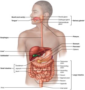
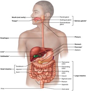
The digestive system is responsible for breaking down food, absorbing nutrients, and eliminating waste. It consists of the alimentary canal and accessory digestive organs, each playing a specific role in digestion and absorption.
Alimentary canal (GI tract): Includes the mouth, pharynx, esophagus, stomach, small intestine, large intestine, and anus.
Accessory digestive organs: Teeth, tongue, gallbladder, salivary glands, liver, and pancreas.
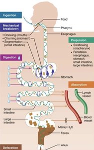
Major functions: Ingestion, mechanical breakdown, propulsion, digestion, absorption, and defecation.
Gastrointestinal Tract Activities
The GI tract performs several coordinated activities to process food:
Ingestion: Intake of food into the mouth.
Mechanical breakdown: Chewing (mouth), churning (stomach), and segmentation (small intestine).
Propulsion: Swallowing and peristalsis move food through the tract.
Digestion: Enzymatic breakdown of food into absorbable molecules.
Absorption: Transport of nutrients from the GI tract into blood or lymph.
Defecation: Elimination of indigestible substances as feces.
Peristalsis and Segmentation
These are two primary motility patterns in the GI tract:
Peristalsis: Adjacent segments of the alimentary canal organs contract and relax, propelling food distally.
Segmentation: Nonadjacent segments contract and relax, mixing food and breaking it down mechanically.
Peritoneum and Mesenteries
Structure and Function
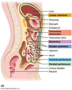
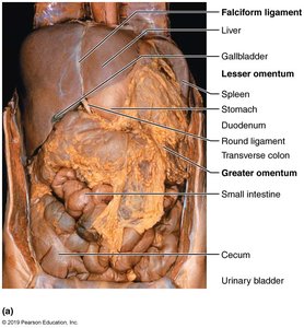
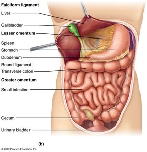
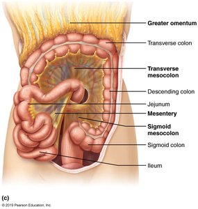
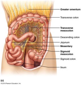
The peritoneum is a serous membrane that lines the abdominopelvic cavity and covers most digestive organs. Mesenteries are double layers of peritoneum that anchor organs and provide pathways for blood vessels, nerves, and lymphatics.
Visceral peritoneum: Covers external surfaces of most digestive organs.
Parietal peritoneum: Lines the body wall.
Retroperitoneal organs: Located outside or posterior to the peritoneum (e.g., cecum, appendix, rectum, parts of colon).
Major folds: Greater omentum, lesser omentum, mesentery, transverse mesocolon, sigmoid mesocolon.
Histology of the Alimentary Canal
Four Common Layers
The alimentary canal is composed of four basic layers, each with distinct functions:
Mucosa: Secretes mucus, absorbs end products of digestion, and protects against infectious disease.
Submucosa: Contains blood vessels, nerves, and lymphatics.
Muscularis externa: Responsible for segmentation and peristalsis; consists of circular and longitudinal muscle layers.
Serosa: Outermost layer; reduces friction between digestive organs and surrounding structures.
Neural Control of the GI Tract
Enteric Nervous System and Reflex Pathways
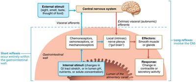
The GI tract is regulated by the enteric nervous system (ENS) and neural reflex pathways. The ENS is sometimes called the "gut brain" and can function independently of the central nervous system.
Short reflexes: Mediated by the ENS; respond to local stimuli within the GI tract.
Long reflexes: Involve the CNS; respond to external stimuli (e.g., sight, smell, taste, thought of food).
Effectors: Smooth muscle and glands change contractile or secretory activity.
Mouth and Associated Organs
Anatomy and Functions
The mouth is the entry point for food and initiates both mechanical and chemical digestion. Associated organs include the oral cavity, labia, buccae, hard and soft palate, tongue, salivary glands, and teeth.
Mechanical digestion: Chewing (mastication) forms a bolus.
Chemical digestion: Salivary amylase and lingual lipase begin breakdown of starch and fats.
Tongue
The tongue is a muscular organ that manipulates food, assists in swallowing, and contains taste buds.
Teeth
Teeth are essential for mechanical digestion. Humans have two sets of teeth:
Primary dentition: 20 deciduous (baby) teeth.
Permanent dentition: 32 teeth that replace primary teeth.
Salivary Glands
Salivary glands produce saliva, which cleanses the mouth, dissolves food chemicals, moistens food, and begins starch digestion.
Serous cells: Produce watery secretion with enzymes and ions.
Mucous cells: Produce mucus.
Saliva composition: Mostly water (97–99.5%), slightly acidic, contains electrolytes, proteins (mucin, lysozyme, IgA), metabolic wastes, and antimicrobial agents.
Pharynx and Esophagus
Deglutition (Swallowing)
Swallowing involves the tongue, soft palate, pharynx, and esophagus. It consists of two phases:
Buccal phase: Voluntary contraction of the tongue.
Pharyngeal-esophageal phase: Involuntary, primarily controlled by the vagus nerve.
Stomach
Gross and Microscopic Anatomy
The stomach is a muscular organ that stores, mixes, and digests food. It has unique modifications:
Muscularis externa: Has an extra oblique layer for churning.
Mucosa: Forms rugae when empty; contains gastric pits leading to gastric glands.
Gastric Secretions
Goblet cells: Secrete mucus.
Parietal cells: Secrete hydrochloric acid (HCl) and intrinsic factor (for vitamin B12 absorption).
Chief cells: Secrete pepsinogen (activated to pepsin by HCl) and lipases.
Enteroendocrine cells: Secrete hormones and paracrines (serotonin, histamine, somatostatin, gastrin).
Small Intestine
Anatomy and Structural Modifications
The small intestine is the primary site for digestion and absorption. It consists of the duodenum, jejunum, and ileum.
Circular folds: Increase surface area.
Villi: Finger-like projections for absorption.
Microvilli: Further increase surface area.
Secretions and Hormonal Control
Brush border enzymes: Peptidase, sucrase, maltase, lactase, intestinal lipase, enterokinase.
Hormones: Gastrin, somatostatin, cholecystokinin (CCK), secretin.
Accessory Digestive Organs
Pancreas
The pancreas has both endocrine and exocrine functions. It produces pancreatic juice containing digestive enzymes and bicarbonate.
Proteases: For protein digestion (secreted in inactive form).
Amylase: For carbohydrate digestion.
Lipases: For lipid digestion.
Nucleases: For nucleic acid digestion.
Liver and Gallbladder
The liver produces bile, which emulsifies fats. The gallbladder stores and concentrates bile.
Bile composition: Bile salts, bilirubin, cholesterol, triglycerides, phospholipids, electrolytes.
Enterohepatic circulation: Recycles bile salts; 95% are reused, 5% excreted.
Absorption in the Small Intestine
Mechanisms and Routes
Absorption occurs primarily in the small intestine, where nutrients, electrolytes, and vitamins are transported into blood or lymph.
Carbohydrates: Absorbed as monosaccharides.
Proteins: Absorbed as amino acids.
Fats: Emulsified by bile, digested by lipases, absorbed as fatty acids and monoglycerides.
Electrolytes and vitamins: Absorbed via specific transport mechanisms.
Large Intestine
Functions and Anatomy
The large intestine absorbs water and electrolytes, forms feces, and houses bacterial flora.
Bacterial flora: Ferment indigestible carbohydrates, synthesize B complex and vitamin K.
Characteristics: Lack villi, contain goblet cells, no digestive enzymes produced.
Defecation Reflex
The defecation reflex is initiated by distension of the rectum, leading to contraction of the rectal walls and relaxation of the internal anal sphincter.
Feces Composition
Water: 75% of feces.
Solids: 25% (includes bacteria, epithelial cells, indigestible materials, mucus, electrolytes).
Bile pigments: Metabolized by bacteria, alter color.
Odor: Produced by bacterial metabolism.
Digestive Organ | Main Function | Key Secretions |
|---|---|---|
Mouth | Mechanical and chemical digestion | Saliva (amylase, lingual lipase) |
Stomach | Mixing, protein digestion | Gastric juice (HCl, pepsinogen, mucus) |
Small Intestine | Digestion, absorption | Brush border enzymes, hormones |
Pancreas | Enzyme production | Pancreatic juice (proteases, amylase, lipase, nucleases) |
Liver | Bile production | Bile (bile salts, bilirubin) |
Large Intestine | Water/electrolyte absorption, feces formation | None (no digestive enzymes) |
Additional info: The notes above expand on brief points with academic context, definitions, and examples to ensure completeness and clarity for exam preparation.
