 Indietro
Study Guide: The Lymphatic System (Anatomy & Physiology)
Guida di studio - Note intelligenti
 Appunti personalizzati basati sui tuoi materiali, ampliati con definizioni chiave, esempi e contesto.
The Lymphatic System Overview
Introduction to the Lymphatic System
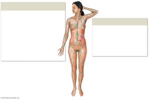
The lymphatic system is a crucial component of the circulatory and immune systems. It is responsible for draining excess interstitial fluid, filtering lymph, and supporting immune functions.
Lymphatic vessels transport lymph, a fluid derived from interstitial fluid.
Lymph nodes filter lymph and remove antigens.
Lymphatic tissues and organs house lymphocytes, which are essential for adaptive immunity.
Lacteals in the digestive system absorb lipids and distribute them slowly into circulation.
Lymphatic Vessels and Lymphatic Drainage
Structure and Function of Lymphatic Vessels
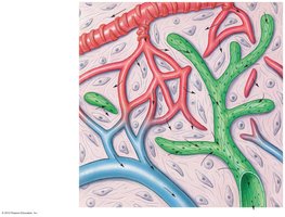
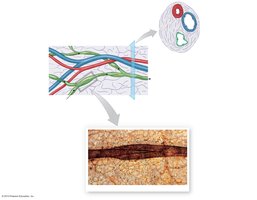
Lymphatic vessels begin as lymphatic capillaries in tissues, which collect excess interstitial fluid. These capillaries drain into larger vessels equipped with valves to ensure one-way flow under low pressure.
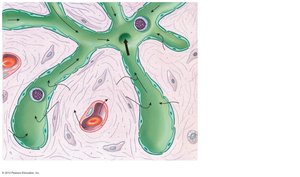
Lymphatic capillaries are lined by endothelial cells with an incomplete or absent basement membrane.
Overlapping endothelial cells act as one-way valves, allowing fluid to enter but not exit.
Lymphatic vessels converge into trunks, and the cisterna chyli collects lipid-rich lymph (chyle) from lacteals.
The final vessels, the right lymphatic duct and thoracic duct, drain lymph into the subclavian veins.
Lymphatic Drainage Pathways
Not all fluid filtered out of capillaries returns directly to the blood; excess fluid enters lymphatic capillaries and is transported through the lymphatic system.
Lymphatic vessels form a network that returns lymph to large veins near the heart.
Drainage occurs via the right lymphatic duct (right upper body) and thoracic duct (rest of the body).
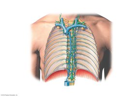
Lymphoid Tissue and Lymphatic Organs
Lymphoid Tissue
Lymphoid tissue consists mainly of fine reticular fibers and houses lymphocytes and macrophages for immune function.
Can be dense (nodules) or diffuse.
Non-encapsulated tissue blends with other organs, while encapsulated organs (e.g., lymph nodes) are distinct.
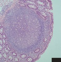
Mucosa-Associated Lymphoid Tissue (MALT) and Tonsils
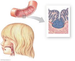
MALT is found in the digestive, respiratory, urinary, and reproductive tracts, providing localized immune defense. Tonsils are specialized lymphoid tissues in the pharynx.
Palatine tonsil is the only tonsil visible on anatomical models.
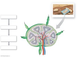
Lymph Nodes
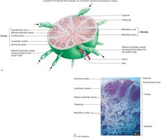
Lymph nodes filter lymph and initiate immune responses.
Remove at least 99% of antigens immediately.
Structural features include afferent vessels (in), efferent vessels (out), outer capsule, nodules in cortex, and diffuse medulla.
The Thymus
Structure and Function
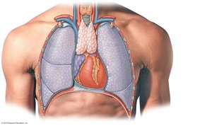
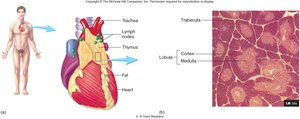
The thymus is a glandular organ in the mediastinum that produces thymosins, hormones promoting T-lymphocyte development. It becomes fibrous and atrophies with age.
Essential for maturation of T-cells (adaptive immunity).
The Spleen
Structure and Functions
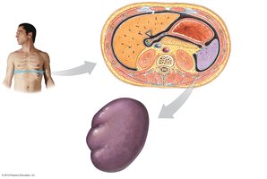
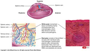
The spleen is the largest mass of lymphatic tissue, with three major functions:
Filters antigens from blood and stimulates immune response.
Destroys defective red blood cells (RBCs).
Acts as a blood reservoir.
Histological Features
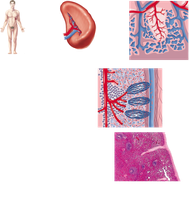
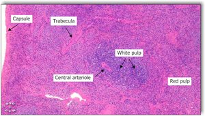
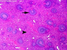
White pulp: Lymphatic tissue surrounding arterial branches, rich in lymphocytes.
Red pulp: Venous sinuses and splenic cords, where blood is processed and defective RBCs are removed.
Summary Table: Major Lymphatic Organs and Their Functions
Organ | Main Function | Key Features |
|---|---|---|
Lymph Node | Filters lymph, initiates immune response | Afferent/efferent vessels, cortex, medulla |
Thymus | Maturation of T-lymphocytes | Produces thymosins, atrophies with age |
Spleen | Filters blood, destroys defective RBCs, blood reservoir | White pulp (lymphatic tissue), red pulp (venous sinuses) |
MALT | Localized immune defense | Diffuse lymphoid tissue in mucosa |
Tonsils | Immune defense in pharynx | Palatine, pharyngeal, lingual tonsils |
Key Terms and Definitions
Lymph: Fluid collected from interstitial spaces, transported by lymphatic vessels.
Lymphocyte: White blood cell involved in adaptive immunity.
Lacteal: Specialized lymphatic capillary in the intestine for lipid absorption.
Cisterna chyli: Dilated lymphatic sac collecting chyle from the intestines.
Thymosin: Hormone produced by the thymus for T-cell development.
White pulp: Lymphatic tissue in the spleen surrounding arteries.
Red pulp: Venous sinuses and splenic cords in the spleen.
Relevant Equations
Fluid Exchange at Capillaries
The movement of fluid between blood and interstitial spaces is governed by Starling's law:
Net filtration pressure (NFP) determines the direction of fluid movement.
Additional info:
Adaptive immunity is mediated by lymphocytes housed in lymphoid tissues.
Lymphatic system is essential for maintaining fluid balance and defending against pathogens.
