 Indietro
Study Guide: The Muscular System and Muscle Physiology
Guida di studio - Note intelligenti
 Appunti personalizzati basati sui tuoi materiali, ampliati con definizioni chiave, esempi e contesto.
The Muscular System
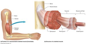
Structure of Skeletal Muscles
Skeletal muscles are composed of long, cylindrical cells called muscle fibers, which are organized into bundles and surrounded by connective tissue layers. This structure allows for efficient force generation and transmission.
Muscle Fiber: The basic unit of skeletal muscle, surrounded by endomysium.
Fascicle: A bundle of 10-100 muscle fibers, surrounded by perimysium.
Epimysium: Connective tissue that surrounds all fascicles in a muscle.
Fascia: The most superficial connective tissue sheath, continuous with epimysium.
Tendons: Attach muscle to bone or other structures.
Blood and Nerve Supply: Skeletal muscles are richly supplied with blood vessels and nerves.
Voluntary Control: Skeletal muscle contraction is under conscious control and requires nervous system stimulation.
Fascicle Patterns and Muscle Shapes
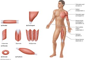
The arrangement of fascicles within a muscle determines its shape and function. Different patterns allow for specialized movements and force generation.
Parallel: Evenly spaced fascicles; e.g., Sartorius.
Convergent: Broad at one end, tapers to a single tendon; e.g., Pectoralis Major.
Circular: Encircles an opening; e.g., Orbicularis Oculi.
Fusiform: Thick in the middle, tapered at ends; e.g., Biceps Brachii.
Pennate: Fascicles attach to tendon at an angle, feather-like appearance.
Unipennate: Fascicles on one side; e.g., Flexor Pollicis Longus.
Bipennate: Fascicles on both sides; e.g., Rectus Femoris.
Multipennate: Several tendons, multiple feather-like fascicle arrangements; e.g., Deltoid.
Naming Muscles
Muscles are named based on their size, location, shape, appearance, position, number of heads, points of attachment, or function.
Size: Major, Minor, Longus, Brevis, Vastus.
Location: Directional terms (Superior, Inferior, Medial, Lateral), regional terms.
Attachment: Named for structures they attach to (e.g., Sternocleidomastoid).
Function: Flexors, Extensors, Levators, Adductors, Abductors.
Depth: Profundus (deep), Superficialis (superficial).
Functional Groups of Muscles
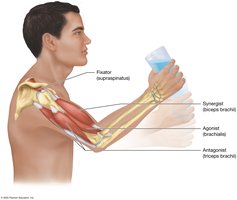
Muscles work in groups to produce coordinated movements. Each group has a specific role in movement.
Agonists (Prime Movers): Provide most of the force for a movement.
Antagonists: Oppose and slow the action of agonists.
Synergists: Assist agonists and guide movement.
Fixators: Stabilize bones and joints, making movement more efficient.
Muscle Origin and Insertion
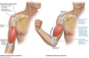
Muscles attach to bones at two points: the origin (more fixed) and the insertion (moves during contraction). The roles can switch depending on the movement.
Origin: Fixed attachment point.
Insertion: Movable attachment point.
Example: Biceps brachii origin is scapula, insertion is radius.
Muscle Physiology
Overview of Muscle Tissue
Muscle tissue generates tension and performs essential functions in the body.
Functions: Movement, posture, joint stabilization, heat generation, regulation of material flow through hollow organs.
Types of Muscle Tissue
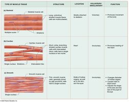
There are three types of muscle tissue, each with unique structure and function.
Type | Structure | Location | Voluntary? | Function |
|---|---|---|---|---|
Striated (Skeletal) | Long, cylindrical, multinucleated, striations | Attached to skeleton | Yes | Movement of body |
Cardiac | Short, branching, single nucleus, striations | Heart | No | Pumping blood |
Smooth | Spindle-shaped, single nucleus, no striations | Walls of hollow organs, eyes | No | Changes diameter, moves material |
Properties of Muscle Cells
Muscle cells possess unique properties that enable their function.
Contractility: Ability to contract and generate force.
Excitability: Ability to respond to stimuli.
Conductivity: Ability to conduct electrical signals.
Elasticity: Ability to return to original length after stretching.
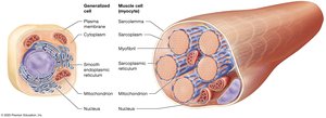
Structure of Muscle Cells
Muscle cells contain specialized organelles for contraction.
Sarcoplasm: Cytoplasm of muscle cell.
Sarcolemma: Plasma membrane of muscle cell.
Myofibrils: Bundles of contractile proteins.
Sarcoplasmic Reticulum (SR): Modified smooth ER, stores calcium ions.
Structure of the Skeletal Muscle Fiber
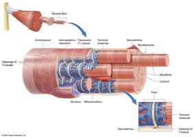
Skeletal muscle fibers are long, cylindrical, multinucleated cells with striations. They contain abundant myofibrils and specialized structures for contraction.
Cell Shape: Long, cylindrical, up to 30 cm.
Striations: Alternating light and dark bands.
Multiple Nuclei: Result from fusion of myoblasts.
T-tubules: Inward extensions of sarcolemma, surround myofibrils.
Triad: T-tubule flanked by two terminal cisternae of SR.

Structure of the Myofibril
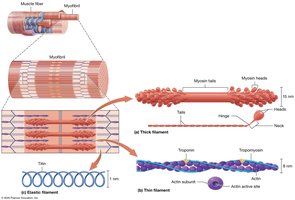
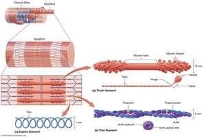
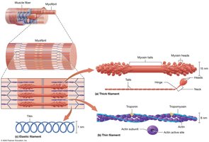
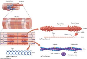
Myofibrils are composed of three types of myofilaments: thick, thin, and elastic. Each type has a specific role in muscle contraction.
Thick Filaments: Made of myosin, with heads that bind actin and ATP.
Thin Filaments: Made of actin, tropomyosin, and troponin.
Elastic Filaments: Made of titin, provide elasticity and stability.
Structure of the Sarcomere
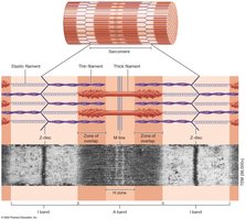
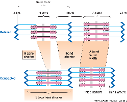
The sarcomere is the functional unit of muscle contraction, defined by the arrangement of myofilaments.
I Band: Light region, only thin filaments.
A Band: Dark region, thick and thin filaments.
H Zone: Middle of A band, only thick filaments.
M Line: Structural proteins holding thick filaments.
Z Disk: Anchors thin and elastic filaments, attaches myofibrils.
The Sliding-Filament Mechanism
Muscle contraction occurs when thin filaments slide past thick filaments, shortening the sarcomere and generating tension.
Myosin heads pull actin filaments toward the M line.
I bands and H zone narrow, A band remains unchanged.
Sarcomere shortens, but filaments do not change length.
The Neuromuscular Junction (NMJ)
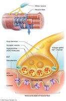
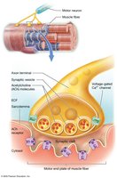
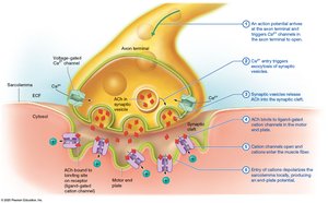
The NMJ is the synapse between a motor neuron and a muscle fiber, essential for muscle stimulation.
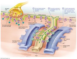
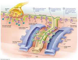
Axon Terminal: Contains synaptic vesicles with acetylcholine (ACh).
Synaptic Cleft: Space between neuron and muscle fiber.
Motor End Plate: Specialized sarcolemma region with ACh receptors.
Skeletal Muscle Contraction: Phases
Muscle contraction occurs in three phases: excitation, excitation-contraction coupling, and contraction.
Excitation: ACh stimulates sarcolemma, generating an action potential.
Excitation-Contraction Coupling: Action potential triggers calcium release from SR.
Contraction: Myosin binds actin, sliding-filament mechanism shortens sarcomere.
Role of Calcium in Muscle Contraction
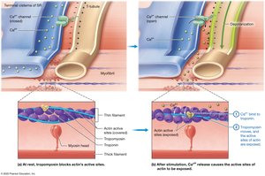
Calcium ions are essential for muscle contraction, binding to troponin and exposing actin's active sites.
Calcium binds troponin, causing tropomyosin to move and expose actin sites.
Myosin binds actin, initiating crossbridge cycle.
The Crossbridge Cycle
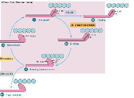
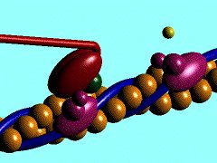
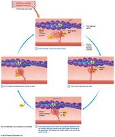
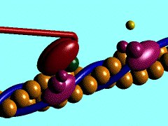
The crossbridge cycle is the sequence of events in muscle contraction, powered by ATP.
ATP hydrolysis cocks myosin head.
Myosin binds actin (crossbridge formation).
Power stroke: Myosin pulls actin toward center, phosphate and ADP released.
Detachment: New ATP binds, myosin releases actin.
Cycle repeats 20–40 times per contraction.
Muscle Relaxation
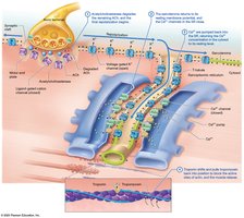
Muscle relaxation occurs when the requirements for contraction are removed.
Acetylcholinesterase degrades ACh, closing cation channels.
Calcium channels in SR close.
Calcium pumps return calcium to SR.
Troponin and tropomyosin block actin sites.
Rigor Mortis
Rigor mortis is the stiffening of muscles after death due to lack of ATP for calcium pumps and myosin detachment.
Calcium remains in cytosol, binding troponin and causing contraction.
Myosin heads cannot detach from actin without ATP.
Stiffness persists until proteins degrade (48–72 hours).

Energy Sources for Skeletal Muscle
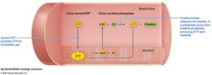
ATP is required for muscle contraction and relaxation. Muscle fibers regenerate ATP through three processes.
Immediate: Creatine phosphate regenerates ATP via creatine kinase.
Glycolytic: Glycolysis in cytosol produces ATP and lactic acid.
Oxidative: Mitochondria use oxygen to produce ATP.
Additional info:
These notes cover the structure and function of skeletal muscle, muscle physiology, and the mechanisms of contraction and relaxation, as outlined in Chapters 9 and 10 of a typical Anatomy & Physiology college course.
