 Indietro
Chapter 22
Guida di studio - Note intelligenti
 Appunti personalizzati basati sui tuoi materiali, ampliati con definizioni chiave, esempi e contesto.
The Respiratory System: Overview
Major Functions and Processes
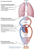
The respiratory system is essential for gas exchange, supplying oxygen to cells for cellular respiration and removing carbon dioxide, a waste product. It works closely with the cardiovascular system to accomplish four key processes collectively known as respiration:
Pulmonary ventilation: Movement of air into and out of the lungs (breathing).
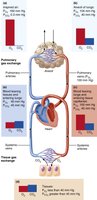
Pulmonary gas exchange: Exchange of gases (O2 and CO2) between the lungs and blood.
Transport of respiratory gases: Movement of gases in the blood between the lungs and tissues.
Tissue gas exchange: Exchange of gases between blood and body tissues.
Functional Anatomy of the Respiratory System
Major Organs and Divisions
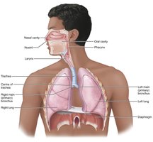
The respiratory system is divided into upper and lower regions, each with specialized structures:
Upper respiratory system: Nose, paranasal sinuses, and pharynx.
Lower respiratory system: Larynx, trachea, bronchi (and branches), lungs, and alveoli.
Respiratory muscles: Classified as part of the muscular system, these include the diaphragm and intercostal muscles.
Surface Anatomy and Skeletal Framework of the Nose
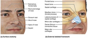
The nose is the only externally visible part of the respiratory system. It provides an airway, moistens and warms air, filters particles, serves as a resonating chamber for speech, and houses olfactory receptors.
Surface features: Root, bridge, dorsum nasi, apex, nostrils (nares).
Skeletal framework: Nasal and frontal bones, maxillary bones, hyaline cartilage (alar and septal cartilages).
Nasal Cavity and Paranasal Sinuses
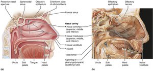
The nasal cavity is divided by the nasal septum and lined with mucous membranes.
Nasal vestibule: Lined with vibrissae (hairs) to filter coarse particles.
Olfactory mucosa: Contains olfactory epithelium for smell.
Respiratory mucosa: Pseudostratified ciliated columnar epithelium with goblet cells; cilia sweep mucus toward the throat.
Nasal conchae: Superior, middle, and inferior conchae increase surface area and enhance turbulent airflow.
Paranasal sinuses: Located in frontal, sphenoid, ethmoid, and maxillary bones; lighten the skull, help warm/moisten air, and drain mucus.
The Pharynx
Regions and Functions
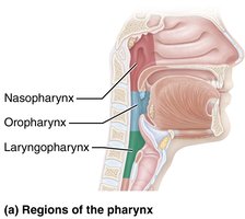
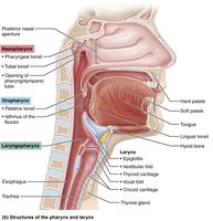
The pharynx connects the nasal cavity to the larynx and mouth to the esophagus. It is divided into three regions:
Nasopharynx: Posterior to nasal cavity; serves only as an airway; lined with pseudostratified ciliated columnar epithelium.
Oropharynx: Posterior to oral cavity; passageway for food and air; lined with stratified squamous epithelium.
Laryngopharynx: Posterior to larynx; passageway for food and air; lined with stratified squamous epithelium.
The Lower Respiratory System
Conducting and Respiratory Zones
Conducting zone: All airways from nose to terminal bronchioles; transport, cleanse, warm, and humidify air.
Respiratory zone: Sites of gas exchange; includes respiratory bronchioles, alveolar ducts, and alveoli.
Larynx (Voice Box)
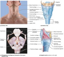
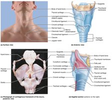
The larynx provides an open airway, routes air and food, and houses vocal folds for sound production.
Framework: Nine cartilages (eight hyaline, one elastic - epiglottis).
Vocal folds: Vibrate to produce sound; glottis is the opening between folds.
Epithelium: Stratified squamous (superior), pseudostratified ciliated columnar (inferior).
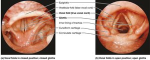
Trachea (Windpipe)
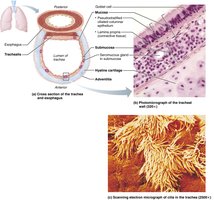
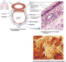
The trachea is a flexible tube supported by C-shaped cartilage rings, lined with ciliated pseudostratified epithelium.
Three layers: Mucosa, submucosa, adventitia.
Trachealis muscle: Contracts during cough to expel mucus.
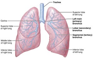
Carina: Last cartilage, marks division into main bronchi.
Bronchi and Subdivisions
Airways branch about 23 times, forming the bronchial tree.
Main bronchi: Right and left, enter lungs at hilum.
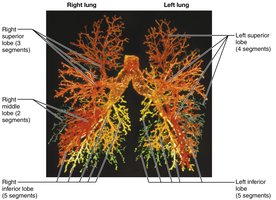
Lobar bronchi: One per lung lobe.
Segmental bronchi: Further divisions.
Bronchioles: Less than 1 mm diameter; terminal bronchioles are less than 0.5 mm.
Respiratory Zone Structures
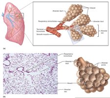
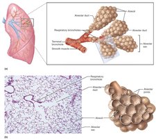
Terminal bronchioles lead to respiratory bronchioles, alveolar ducts, and alveolar sacs.
Alveoli: Sites of gas exchange; millions in each lung.
Alveoli and Respiratory Membrane
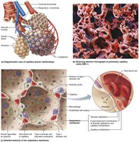
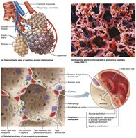
The respiratory membrane is a thin blood-air barrier for gas exchange.
Alveolar wall: Simple squamous epithelium (type I cells).
Type II cells: Secrete surfactant and antimicrobial proteins.
Alveolar macrophages: Clean inner surfaces.
Alveolar pores: Equalize air pressure and provide alternate routes.
Gross Anatomy of the Lungs
Lung Structure and Lobes
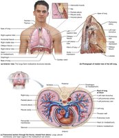
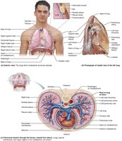
Each lung is surrounded by pleurae and connected to the mediastinum.
Left lung: Superior and inferior lobes, separated by oblique fissure; smaller due to heart position.
Right lung: Superior, middle, and inferior lobes; separated by horizontal and oblique fissures.
Bronchopulmonary segments: 10 in right, 8–10 in left; each served by its own artery, vein, and bronchus.
Lobules: Smallest subdivisions, hexagonal shape.
Blood Supply and Innervation
Pulmonary circulation: Pulmonary arteries carry deoxygenated blood to lungs; veins return oxygenated blood to heart.
Bronchial circulation: Bronchial arteries supply lung tissue (except alveoli).
Innervation: Parasympathetic (bronchoconstriction), sympathetic (bronchodilation), and visceral sensory fibers.
Pleurae
Parietal pleura: Lines thoracic wall and diaphragm.
Visceral pleura: Covers external lung surface.
Pleural fluid: Lubricates and creates surface tension to keep lungs attached to thoracic wall.
Pulmonary Ventilation: Mechanics of Breathing
Pressure Relationships
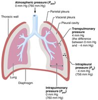
Atmospheric pressure: Pressure exerted by air surrounding the body (760 mm Hg at sea level).
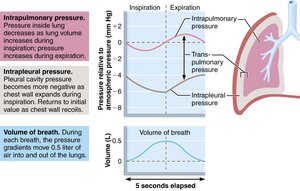
Intrapulmonary pressure: Pressure in alveoli; fluctuates during breathing, equalizes with atmospheric pressure.
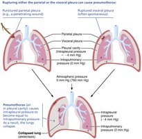
Intrapleural pressure: Pressure in pleural cavity; always negative relative to intrapulmonary pressure.
Transpulmonary pressure: Difference between intrapulmonary and intrapleural pressures; keeps lungs open.
Boyle’s Law and Breathing
Boyle’s law describes the relationship between pressure and volume of a gas:
Pressure (P) varies inversely with volume (V):
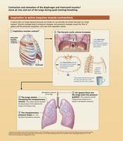
Inspiration: Diaphragm and external intercostals contract, increasing thoracic volume and decreasing pressure, causing air to flow in.
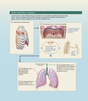
Expiration: Muscles relax, thoracic volume decreases, pressure increases, air flows out.
Physical Factors Influencing Pulmonary Ventilation
Airway Resistance
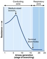
Friction in airways is the major source of resistance.
Flow (F) is directly proportional to pressure difference and inversely proportional to resistance:
Greatest resistance occurs in medium-sized bronchi.
Alveolar Surface Tension
Surface tension at the gas-liquid interface tends to collapse alveoli.
Surfactant (produced by type II alveolar cells) reduces surface tension, preventing collapse.
Lung Compliance
Compliance is the measure of lung expansion per unit pressure change:
High compliance means easier ventilation; affected by tissue distensibility and surfactant levels.
Pulmonary Volumes and Capacities
Key Volumes
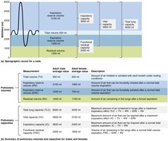
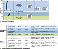
Tidal volume (TV): Air moved in/out with each breath (~500 mL).
Inspiratory reserve volume (IRV): Air forcibly inspired beyond TV (2100–3200 mL).
Expiratory reserve volume (ERV): Air forcibly expired beyond TV (1000–1200 mL).
Residual volume (RV): Air remaining in lungs after forced expiration.
Key Capacities
Inspiratory capacity (IC): TV + IRV
Functional residual capacity (FRC): RV + ERV
Vital capacity (VC): TV + IRV + ERV
Total lung capacity (TLC): TV + IRV + ERV + RV
Gas Exchange: Diffusion Between Blood, Lungs, and Tissues
Basic Properties of Gases
Dalton’s law: Total pressure of a gas mixture is the sum of partial pressures of individual gases.
Henry’s law: The amount of gas dissolved in a liquid is proportional to its partial pressure and solubility.
Pulmonary Gas Exchange
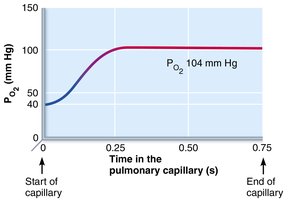
Driven by partial pressure gradients and gas solubilities.
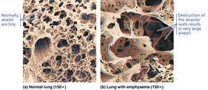
Respiratory membrane thickness and surface area affect exchange efficiency.
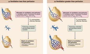
Ventilation-perfusion coupling matches air flow to blood flow for optimal exchange.
Oxygen Transport
Hemoglobin and Oxygen Saturation
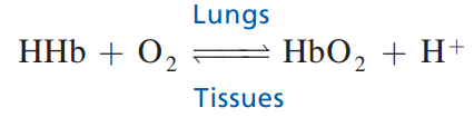
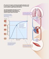
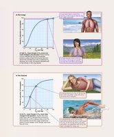
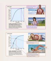
98.5% of O2 is bound to hemoglobin (Hb) in RBCs; 1.5% dissolved in plasma.
Each Hb molecule can carry four O2 molecules.
Oxygen loading/unloading is influenced by partial pressure, temperature, pH, and BPG levels.
Carbon Dioxide Transport
Forms of CO2 Transport
Dissolved in plasma (7–10%).
Bound to hemoglobin as carbaminohemoglobin (20%).
As bicarbonate ions in plasma (70%).
Neural Control of Respiration
Respiratory Centers
Medullary centers: Ventral (VRG) and dorsal (DRG) groups generate rhythm and integrate input.
Pontine centers: Smooth transitions between inspiration and expiration.
Chemical and Neural Influences
Central and peripheral chemoreceptors monitor CO2, O2, and pH.
CO2 is the most powerful respiratory stimulant.
Higher brain centers, pulmonary irritant reflexes, and inflation reflex also affect breathing.
Clinical Applications and Disorders
Common Disorders
Rhinitis: Inflammation of nasal mucosa.
Laryngitis: Inflammation of vocal folds.
Pleurisy: Inflammation of pleurae.
Atelectasis: Lung collapse due to plugged bronchioles or pneumothorax.
Infant respiratory distress syndrome (IRDS): Lack of surfactant in premature infants.
Chronic Obstructive Pulmonary Disease (COPD): Includes emphysema and chronic bronchitis; characterized by decreased ability to force air out.
Asthma: Acute, reversible airway obstruction due to inflammation.
Tuberculosis: Infectious disease caused by Mycobacterium tuberculosis.
Lung cancer: Leading cause of cancer deaths; most cases due to smoking.
Sleep apnea: Temporary cessation of breathing during sleep.
Cystic fibrosis: Genetic disorder causing thick mucus and respiratory infections.
Developmental Aspects
Embryonic Development
Upper respiratory structures develop first; olfactory placodes form nasal cavities by week 4.
Laryngotracheal bud forms tracheal lining and mucosae of bronchi and alveoli by week 5.
By week 28, most premature babies can breathe independently.
At birth, respiratory centers activate, alveoli inflate, and lungs begin functioning.
Summary Table: Upper and Lower Respiratory System
Region | Main Structures | Functions |
|---|---|---|
Upper Respiratory | Nose, paranasal sinuses, pharynx | Airway, filtration, humidification, olfaction, speech resonance |
Lower Respiratory | Larynx, trachea, bronchi, lungs, alveoli | Airway, sound production, gas exchange |
Summary Table: Pulmonary Volumes and Capacities
Volume/Capacity | Definition | Average Value (Adult Male) |
|---|---|---|
Tidal Volume (TV) | Amount of air inhaled/exhaled with each breath | 500 mL |
Inspiratory Reserve Volume (IRV) | Amount of air forcibly inspired beyond TV | 3100 mL |
Expiratory Reserve Volume (ERV) | Amount of air forcibly expired beyond TV | 1200 mL |
Residual Volume (RV) | Amount of air remaining after forced expiration | 1200 mL |
Vital Capacity (VC) | TV + IRV + ERV | 4800 mL |
Total Lung Capacity (TLC) | TV + IRV + ERV + RV | 6000 mL |
Key Equations
Boyle’s Law:
Flow Equation:
Lung Compliance:
Additional info:
Some clinical and developmental details were expanded for clarity and completeness.
Tables were recreated to summarize key comparisons and definitions.
