 Indietro
Study Guide: The Respiratory System (Anatomy & Physiology)
Guida di studio - Note intelligenti
 Appunti personalizzati basati sui tuoi materiali, ampliati con definizioni chiave, esempi e contesto.
The Respiratory System
Overview and Importance
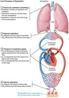
The respiratory system is essential for gas exchange, providing oxygen to the body and removing carbon dioxide. It works closely with the circulatory system and also plays roles in olfaction and speech.
Gas Exchange: Oxygen is brought into the body and carbon dioxide is expelled.
Coupling with Circulatory System: Blood transports gases between lungs and tissues.
Other Functions: Olfaction (smell) and speech production.
Major Organs of the Respiratory System
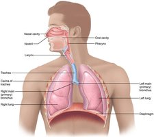
The respiratory system is divided into upper and lower regions, each with distinct anatomical structures and functions.
Upper Respiratory: Nose, nasal cavity, paranasal sinuses, pharynx
Lower Respiratory: Larynx, trachea, bronchi and branches, lungs, alveoli
Upper Respiratory System
The Nose and Paranasal Sinuses
The nose is the only external part of the respiratory system and serves several important functions.
Airway for Respiration: Entry point for air.
Moistens and Warms Air: Prepares air for the lungs.
Filters and Cleans Air: Removes particles and pathogens.
Resonating Chamber: Aids in speech.
Olfactory Receptors: Enables sense of smell.
Nasal Conchae
Function: Filter, heat, and moisten air during inhalation; reclaim heat and moisture during exhalation.
Paranasal Sinuses
Location: Frontal, sphenoid, ethmoid, and maxillary bones.
Functions: Lighten skull, secrete mucus, help warm and moisten air.
Clinical Note: Rhinitis and Sinusitis
Rhinitis: Inflammation of nasal mucosa; can spread to throat, chest, tear ducts, and sinuses.
Sinusitis: Blockage and inflammation of sinuses due to infection.
The Pharynx
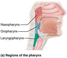
The pharynx is a muscular tube connecting the nasal cavity and mouth to the larynx and esophagus. It is divided into three regions:
Nasopharynx
Oropharynx
Laryngopharynx
Clinical Note: Adenoids
Swollen Adenoids: Can block air passage, forcing mouth breathing and affecting speech and sleep.
Lower Respiratory System
Zones of the Lower Respiratory System
Respiratory Zone: Site of gas exchange; includes respiratory bronchioles, alveolar ducts, and alveoli.
Conducting Zone: Conduits for air transport; cleanses, warms, and humidifies air.
The Larynx
The larynx, or voice box, is attached to the hyoid bone and extends from the 3rd to 6th cervical vertebra. It serves three main functions:
Provides Patent Airway: Keeps airway open.
Routes Air and Food: Directs air to lungs and food to esophagus.
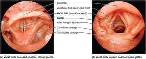
Voice Production: Houses vocal folds for sound generation.

Epiglottis
Structure: Elastic cartilage covered in mucosa with taste buds.
Function: Covers laryngeal inlet during swallowing.
Clinical Note: Laryngitis
Laryngitis: Inflammation of vocal folds, often due to infection or overuse, causing voice changes.
The Trachea
The trachea, or windpipe, is a flexible tube extending from the larynx into the mediastinum, dividing into two main bronchi.
Length: About 4 inches long, 3/4 inch in diameter.
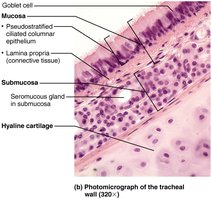
Wall Layers: Mucosa (ciliated pseudostratified epithelium with goblet cells), submucosa, and hyaline cartilage.

Trachealis Muscle
Function: Contracts during coughing to expel mucus.
Carina
Location: Last tracheal cartilage at the branching point.
Function: Highly sensitive; triggers violent coughing if contacted by foreign objects.
Clinical Note: Heimlich Maneuver
Procedure: Uses air in lungs to expel obstructing food.
The Bronchi and Subdivisions
The bronchial tree undergoes 23 orders of branching, transitioning from conducting zone to respiratory zone structures.
Conducting Zone: Bronchi and bronchioles transport air.
Respiratory Zone: Terminal bronchioles lead to respiratory bronchioles, alveolar ducts, and alveolar sacs.
Respiratory Membrane and Alveoli
The respiratory membrane is the site of gas exchange, consisting of alveolar and capillary walls with fused basement membranes.
Thickness: Very thin (~0.5 μm), allowing rapid diffusion.
Alveolar Walls: Type I alveolar cells (squamous epithelium), Type II alveolar cells (secrete surfactant and antimicrobial proteins).
Lungs and Pleurae
Gross Anatomy of the Lungs
The lungs are divided into lobes and bronchopulmonary segments, each served by its own artery, vein, and bronchus.
Right Lung: 10 segments
Left Lung: 8–10 segments
Lobules: Smallest subdivisions, served by bronchioles.
Composition: Mostly alveoli, with elastic connective tissue (stroma).
The Pleurae
The pleurae are thin, double-layered serosal membranes dividing the thoracic cavity into two pleural compartments and the mediastinum.
Pleural Fluid: Fills pleural cavity, providing lubrication and surface tension for lung expansion and recoil.
Clinical Note: Pleurisy and Pleural Effusion
Pleurisy: Inflammation of pleurae, often due to pneumonia, causing pain and friction.
Pleural Effusion: Excess fluid accumulation in pleural cavity, hindering breathing.
Other Fluids: Blood (from damaged vessels), blood filtrate (from heart failure).
Summary Table: Major Structures and Functions of the Respiratory System
Structure | Main Function |
|---|---|
Nose & Nasal Cavity | Airway, moistens/warms/filters air, olfaction, speech |
Pharynx | Connects nasal cavity/mouth to larynx/esophagus |
Larynx | Airway, routes air/food, voice production |
Trachea | Air passage, cleanses/warms/humidifies air |
Bronchi/Bronchioles | Air transport, branching to alveoli |
Alveoli | Gas exchange |
Lungs | Contain alveoli, elastic tissue for expansion |
Pleurae | Lubrication, compartmentalization |
Key Equations
Gas exchange across the respiratory membrane occurs by simple diffusion, governed by Fick's Law:
J: Rate of diffusion
D: Diffusion coefficient
ΔC: Concentration gradient
Δx: Thickness of membrane
Clinical Applications
Rhinitis/Sinusitis: Infections can spread through connected mucosa.
Laryngitis: Voice changes due to inflammation.
Heimlich Maneuver: Emergency procedure for airway obstruction.
Pleurisy/Pleural Effusion: Breathing difficulties due to inflammation or fluid accumulation.
Summary
The respiratory system is vital for gas exchange, voice production, and olfaction. Its anatomy and physiology are closely integrated with the circulatory system, and understanding its structure and function is essential for recognizing clinical conditions and their impact on health.
