 Indietro
Study Guide: The Respiratory System (Anatomy & Physiology)
Guida di studio - Note intelligenti
 Appunti personalizzati basati sui tuoi materiali, ampliati con definizioni chiave, esempi e contesto.
The Respiratory System
Overview of Respiration
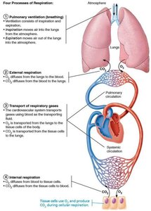
The respiratory system is responsible for providing oxygen (O2) to body tissues and removing carbon dioxide (CO2). Respiration involves several processes that ensure gas exchange and transport throughout the body.
Pulmonary Ventilation: Movement of air into and out of the lungs (inspiration and expiration).
External Respiration: Exchange of gases between lungs and blood; O2 diffuses into blood, CO2 diffuses into alveoli.
Gas Transport: Blood carries O2 and CO2 between lungs and tissues.
Internal Respiration: Exchange of gases between blood and tissues; O2 diffuses into tissues, CO2 diffuses into blood.
Cellular Respiration: Cells use O2 to produce ATP, generating CO2 as a waste product.
Anatomy of the Respiratory System
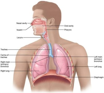
The respiratory system is divided into upper and lower regions, each with specialized structures for air conduction and gas exchange.
Upper Respiratory System
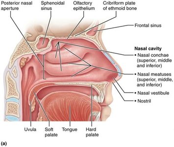
Nares (Nostrils): Main external openings for air entry.
Nasal Cavity: Warms, humidifies, and filters air; contains olfactory mucosa for smell and conchae to increase surface area.
Paranasal Sinuses: Reduce skull weight and contribute to sound resonance.
Pharynx
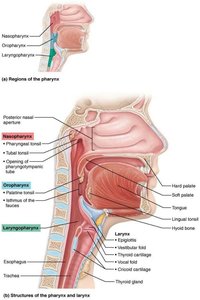
The pharynx is a muscular tube that serves as a passageway for both air and food.
Nasopharynx: Air passageway; contains pharyngeal and tubal tonsils.
Oropharynx: Passageway for food and air; contains palatine and lingual tonsils.
Laryngopharynx: Passageway for food and air; extends to the esophagus.
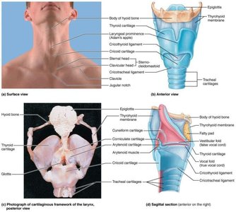
Larynx
The larynx, or voice box, is involved in sound production and air passage. It is composed of several cartilages, including the epiglottis, which prevents food from entering the airway during swallowing.
Epiglottis: Elastic cartilage covering the glottis during swallowing.
Vocal Cords: Produce sound; tension determines pitch.
Lower Respiratory System
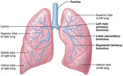
Trachea: Windpipe; reinforced by hyaline cartilage rings and lined with ciliated epithelium.
Bronchial Tree: Branching system of bronchi and bronchioles; cartilage decreases and smooth muscle increases as tubes become smaller.
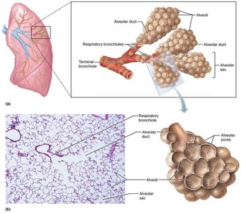
Conducting Zone: No gas exchange; extends from nares to terminal bronchioles.
Respiratory Zone: Gas exchange occurs in respiratory bronchioles, alveolar ducts, and alveoli.
Lungs and Pleurae
Lungs: Located in the pleural cavity; right lung has three lobes, left lung has two.
Pulmonary Circulation: Oxygenates blood and returns it to the heart.
Bronchial Circulation: Supplies oxygenated blood to lung tissues.
Pleurae: Serous membranes with visceral and parietal layers; pleural fluid reduces friction.
Respiratory Physiology: Pressure and Ventilation
Gas movement in the lungs is governed by pressure differences and physical laws.
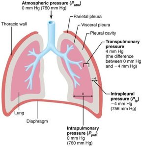
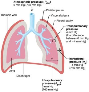
Atmospheric Pressure (Patm): 760 mm Hg at sea level.
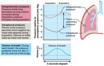
Intrapulmonary Pressure (Ppul): Pressure within the alveoli; equalizes with Patm when airway is open.
Intrapleural Pressure (Pip): 4 mm Hg less than Ppul; keeps lungs expanded.
Transpulmonary Pressure: Difference between Ppul and Pip; maintains lung inflation.
Boyle’s Law: ; gas pressure varies inversely with volume.
Mechanics of Breathing
Inspiration: Active process; diaphragm and external intercostals contract, increasing thoracic volume and decreasing pressure.
Expiration: Passive process; muscles relax, thoracic volume decreases, pressure increases.
Forced Expiration: Involves internal intercostals and abdominal muscles.
Physical Factors: Airway resistance, alveolar surface tension (countered by surfactant), and lung compliance affect ventilation.
Respiratory Volumes and Capacities
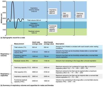
Respiratory volumes and capacities are important for assessing lung function. Dead space refers to air that does not participate in gas exchange.
Anatomical Dead Space: Air left in conducting zone.
Alveolar Dead Space: Air in blocked or collapsed alveoli.
Total Dead Space: Sum of anatomical and alveolar dead space.
Alveolar Ventilation: Volume of air reaching alveoli per minute.
Gas Exchange and Transport
Gas Exchange Principles
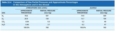
Dalton’s Law: Total pressure of a gas mixture equals the sum of partial pressures of individual gases.
Henry’s Law: Gases dissolve in liquids in proportion to their partial pressures.
Alveolar Gas Composition: Varies with breathing patterns; deep rapid breathing increases O2 and decreases CO2.
Gas | Atmosphere % | Atmosphere Partial Pressure (mm Hg) | Alveoli % | Alveoli Partial Pressure (mm Hg) |
|---|---|---|---|---|
N2 | 78.6 | 597 | 74.9 | 569 |
O2 | 20.9 | 159 | 13.7 | 104 |
CO2 | 0.04 | 0.3 | 5.2 | 40 |
H2O | 0.46 | 3.7 | 6.2 | 47 |
Total | 100.0 | 760 | 100.0 | 760 |
External Respiration
O2 and CO2 diffuse down their partial pressure gradients in opposite directions.
Membrane thickness and surface area affect diffusion rates.
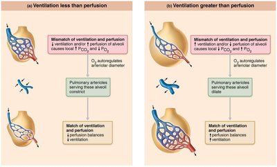
Ventilation-perfusion coupling ensures optimal gas exchange.
Gas Transport
Oxygen Transport: 98.5% carried by hemoglobin as oxyhemoglobin; affinity increases with each O2 bound.
Bohr Effect: High temperature, CO2, and low pH enhance O2 unloading.
CO2 Transport: 7-10% dissolved in plasma, 20% bound to hemoglobin (carbaminohemoglobin), 70% as bicarbonate.
Haldane Effect: Lower O2 increases CO2 carrying capacity.
Internal Respiration
O2 diffuses from blood to tissues; CO2 diffuses from tissues to blood.
Hypoxia: Inadequate O2 delivery; can be anemic, ischemic, histotoxic, or hypoxemic.
CO Poisoning: Hemoglobin binds CO 200x more strongly than O2.
Control of Respiration
Neural Mechanisms: Medullary respiratory centers stimulate breathing muscles.
Eupnea: Normal respiratory rate (12 breaths/min).
Chemical Factors: Chemoreceptors respond to CO2, O2, and pH.
CO2: Most important stimulus; high levels increase rate and depth.
O2: Less important; large reserve and tolerance to low levels.
Arterial pH: Low pH usually due to high CO2.
Higher Brain Centers: Emotional states and voluntary control affect breathing.
Pulmonary Irritants: Sneezing, coughing, etc.
Inflation Reflex: Prevents over-inflation of lungs.
Exercise and Altitude: Exercise increases rate and depth; high altitude leads to acclimatization.
Homeostatic Imbalances
Chronic Obstructive Pulmonary Diseases (COPD): Includes emphysema (damaged alveoli) and chronic bronchitis (excess mucus).
Asthma: Inflammation and constriction of bronchioles.
Tuberculosis (TB): Bacterial infection causing lung nodules.
Lung Cancer: Leading cause of cancer death; often linked to smoking.
Sleep Apnea: Cessation of breathing during sleep; can be obstructive or central.
Example: In asthma, airway resistance increases, making breathing more difficult. In emphysema, alveolar surface area is reduced, impairing gas exchange.
Additional info: This guide expands on brief lecture notes to provide definitions, examples, and context for each major topic in respiratory anatomy and physiology.
