 Indietro
Study Guide: The Respiratory System (Anatomy & Physiology, Chapter 22)
Guida di studio - Note intelligenti
 Appunti personalizzati basati sui tuoi materiali, ampliati con definizioni chiave, esempi e contesto.
The Respiratory System: Overview
Major Functions of the Respiratory System
The respiratory system is essential for gas exchange, supplying oxygen to the body for cellular respiration and removing carbon dioxide, a waste product. It also assists in pH homeostasis, is closely coupled with the circulatory system, and plays roles in olfaction and speech.
Oxygen Supply: Delivers O2 to tissues for cellular respiration.
Carbon Dioxide Removal: Eliminates CO2 produced by cells.
pH Regulation: Helps maintain acid-base balance.
Other Functions: Olfaction (smell) and speech production.
Gross Anatomy of the Respiratory Tract
Major Respiratory Organs and Their Relations
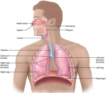
The respiratory tract is divided into upper and lower regions, each with distinct anatomical structures that facilitate air movement and gas exchange.
Upper Respiratory Tract: Includes the nose, nasal cavity, and pharynx.
Lower Respiratory Tract: Includes the larynx, trachea, bronchi, and lungs.
The External Nose
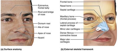
The nose is the primary entryway for air and is composed of surface anatomy and an external skeletal framework.
Surface Anatomy: Features include the dorsum nasi, apex, nostrils, and ala.
Skeletal Framework: Composed of nasal bones, septal cartilage, maxillary bone, and various cartilages.
The Nasal Cavity
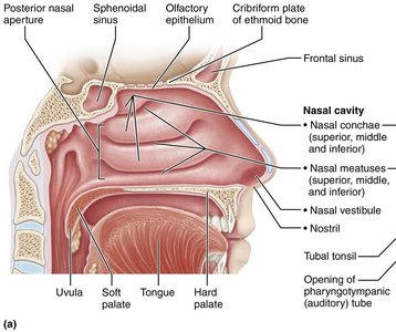
The nasal cavity filters, warms, and moistens incoming air. It contains conchae, meatuses, and is lined with mucosa.
Nasal Conchae: Superior, middle, and inferior structures that increase surface area.
Nasal Meatuses: Passageways beneath each concha.
Other Features: Vestibule, olfactory epithelium, and sinuses.
Paranasal Sinuses
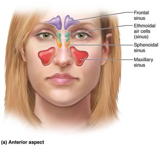
Paranasal sinuses are air-filled spaces that lighten the skull and help warm and moisten air.
Frontal Sinus
Ethmoidal Air Cells
Sphenoidal Sinus
Maxillary Sinus
Pharynx
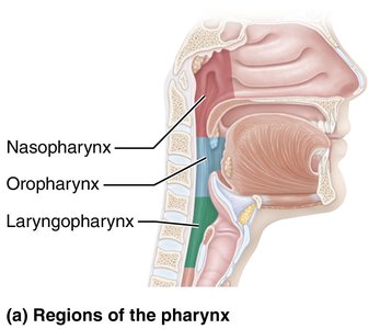
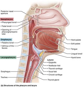
The pharynx is a muscular tube that connects the nasal cavity to the larynx and esophagus. It is divided into three regions: nasopharynx, oropharynx, and laryngopharynx.
Nasopharynx: Posterior to nasal cavity; contains pharyngeal tonsil.
Oropharynx: Posterior to oral cavity; contains palatine and lingual tonsils.
Laryngopharynx: Posterior to larynx; passageway for food and air.
Larynx
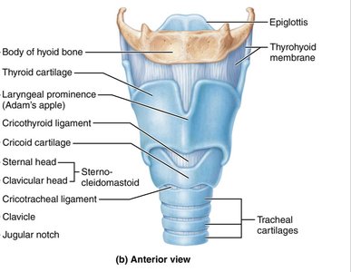
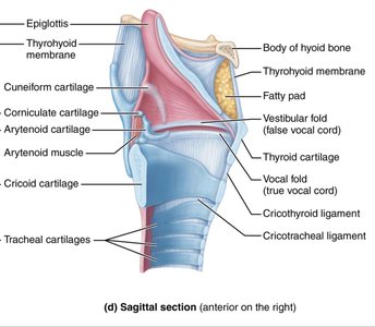
The larynx is the voice box, responsible for sound production and protecting the airway during swallowing. It is composed of cartilages and ligaments.
Thyroid Cartilage: Forms the Adam's apple.
Cricoid Cartilage: Provides support.
Epiglottis: Prevents food from entering the trachea.
Vocal Folds: Vibrate to produce sound.
Glottis and Vocal Folds
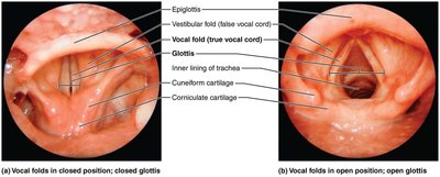
The glottis is the opening between the vocal folds. The folds vibrate to produce sound, and their position determines pitch.
High Pitch: Narrow glottis.
Low Pitch: Wide glottis.
Tissue Composition of the Tracheal Wall
Tracheal Wall Structure
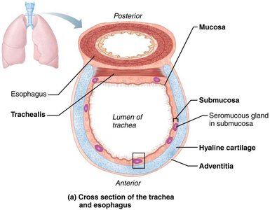
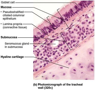
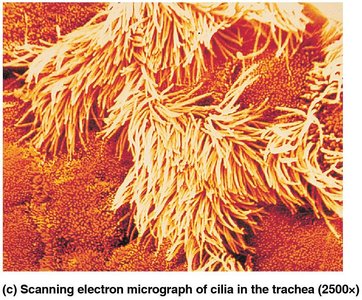
The trachea is supported by cartilaginous rings and lined with mucosa containing ciliated epithelium and goblet cells.
Mucosa: Pseudostratified ciliated columnar epithelium.
Submucosa: Contains seromucous glands.
Hyaline Cartilage: Provides structural support.
Adventitia: Outermost connective tissue layer.
Conducting and Respiratory Zones
Conducting Zone Passages
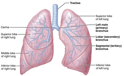
The conducting zone includes all structures that transport air to the respiratory zone, such as the trachea, bronchi, and bronchioles.
Trachea
Primary Bronchi
Secondary (Lobar) Bronchi
Tertiary (Segmental) Bronchi
Respiratory Zone Structures
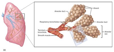
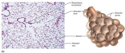
The respiratory zone is where gas exchange occurs, including respiratory bronchioles, alveolar ducts, and alveoli.
Respiratory Bronchioles
Alveolar Ducts
Alveolar Sacs
Alveoli: Main site of gas exchange.
Alveoli and the Respiratory Membrane
Structure and Function
The respiratory membrane is the blood-air barrier, consisting of alveolar and capillary walls with fused basement membranes. It is very thin, allowing efficient gas exchange by simple diffusion.
Type I Alveolar Cells: Squamous epithelium for gas exchange.
Type II Alveolar Cells: Secrete surfactant and antimicrobial proteins.
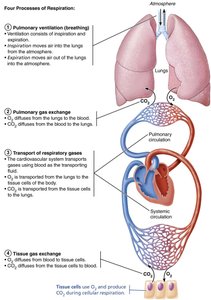
Respiration: The Four Processes
Overview of Respiratory Processes
Respiration involves four processes, divided between the respiratory and circulatory systems:
Pulmonary Ventilation: Movement of air into and out of lungs.
External Respiration: Exchange of O2 and CO2 between lungs and blood.
Transport of Gases: Movement of O2 and CO2 in blood.
Internal Respiration: Exchange of O2 and CO2 between blood and tissues.
Mechanics of Breathing
Pulmonary Ventilation
Pulmonary ventilation consists of inspiration and expiration, driven by changes in thoracic cavity volume and pressure.
Inspiration: Active process involving diaphragm and external intercostals.
Expiration: Usually passive, but can be active during forced breathing.
Boyle's Law: Pressure and volume of a gas are inversely related:
Atmospheric Pressure: 760 mm Hg at sea level.
Physical Factors Influencing Pulmonary Ventilation
Airway Resistance
Airway resistance affects airflow, determined by the diameter of respiratory passageways.
Relationship:
Pressure Gradient: Difference between atmospheric and alveolar pressure.
Alveolar Surface Tension
Surface tension resists expansion of alveoli. Surfactant reduces surface tension, preventing alveolar collapse.
Surfactant: Lipid-protein complex secreted by type II alveolar cells.
Lung Compliance
Lung compliance measures the ease of lung expansion. High compliance means easier expansion; low compliance can result from fibrosis, reduced surfactant, or decreased thoracic flexibility.
Distensibility: Stretchability of lung tissue.
Surfactant: Decreases surface tension, increasing compliance.
Gas Exchange
External and Internal Respiration
Gas exchange occurs in the lungs (external respiration) and tissues (internal respiration), governed by gas laws and partial pressures.
Dalton's Law: Total pressure of a gas mixture equals the sum of partial pressures.
Henry's Law: Gas dissolves in liquid in proportion to its partial pressure.
Oxygen and Carbon Dioxide Transport
Oxygen Transport
Oxygen is carried in blood dissolved in plasma (1.5%) and bound to hemoglobin (98.5%).
Oxyhemoglobin: Hemoglobin bound to O2.
Carbon Dioxide Transport
CO2 is transported dissolved in plasma (7-10%), bound to hemoglobin (20%), and as bicarbonate ions (70%).
Bicarbonate Formation:
Oxygen-Hemoglobin Dissociation Curve
The curve shows the relationship between partial pressure of oxygen and hemoglobin saturation. Factors such as temperature, pH, and PCO2 affect oxygen release.
Bohr Effect: Lower pH increases O2 release.
Haldane Effect: Higher PCO2 increases O2 release.
Control of Respiration
Local Regulation
Ventilation-perfusion coupling ensures efficient gas exchange by matching airflow and blood flow in the lungs.
Lung Perfusion: Blood flow to alveoli.
Alveolar Ventilation: Airflow to alveoli.
Neural Regulation
Respiratory centers in the medulla regulate breathing rate and depth. The ventral and dorsal respiratory groups are key clusters of neurons.
Ventral Respiratory Group: Controls rhythm.
Dorsal Respiratory Group: Integrates sensory input.
Clinical Considerations
Lung Cancer
Lung cancer is the leading cause of cancer deaths, with smoking as the primary risk factor. Types include adenocarcinoma, squamous cell carcinoma, and small cell carcinoma.
Treatment: Surgery, radiation, chemotherapy, and emerging therapies.
Impact of Smoking
Smoking damages cilia, leading to mucus accumulation and increased risk of COPD. Ciliary function can recover after cessation.
Smoker's Cough: Result of lost ciliary function.
COPD: Chronic obstructive pulmonary disease, often linked to smoking.
