 Indietro
Study Guide: The Skeletal System (Essentials of Human Anatomy & Physiology, Chapter 5)
Guida di studio - Note intelligenti
 Appunti personalizzati basati sui tuoi materiali, ampliati con definizioni chiave, esempi e contesto.
The Skeletal System
Overview and Components
The skeletal system is a fundamental organ system in human anatomy, providing structure, protection, and facilitating movement. It consists of bones, joints, cartilages, and ligaments, and is divided into two main subdivisions: the axial skeleton and the appendicular skeleton.
Bones: The rigid framework of the body.
Joints: Articulations where two or more bones meet.
Cartilages: Flexible connective tissue found in joints and other areas.
Ligaments: Dense connective tissue connecting bones.
Subdivisions of the Skeleton
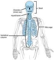
Axial Skeleton: Includes the skull, vertebral column, and rib cage. Provides central support and protection for vital organs.
Appendicular Skeleton: Comprises the limbs, shoulder girdle, and pelvic girdle. Facilitates movement and interaction with the environment.
Functions of the Skeletal System
Support: Maintains body shape and posture.
Protection: Shields soft organs (e.g., skull protects brain, rib cage protects thoracic organs).
Movement: Muscles attach to bones, acting as levers.
Storage: Stores minerals (calcium, phosphorus) and fats (yellow marrow).
Hematopoiesis: Formation of blood cells in red marrow.
Classification of Bones
Types of Osseous Tissue
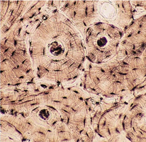
Compact Bone: Dense, smooth, and homogeneous tissue.
Spongy Bone: Contains trabeculae (needlelike structures) and open spaces.
Bone Shapes
Long Bones: Longer than wide, mostly compact bone (e.g., femur, humerus).
Short Bones: Cube-shaped, mostly spongy bone (e.g., carpals, tarsals, patella).
Flat Bones: Thin, flattened, usually curved; two layers of compact bone sandwiching spongy bone (e.g., skull, ribs, sternum).
Irregular Bones: Complex shapes not fitting other categories (e.g., vertebrae, hip bones).
Tissues of the Skeletal System
Types of Connective Tissue
Bones are organs composed of multiple tissue types, including osseous tissue, cartilage, adipose tissue, and blood.
Bone Tissue: Provides structural support and protection.
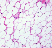
Adipose Tissue: Stores fat in yellow marrow.
Hyaline Cartilage: Found in joints, articular surfaces, and epiphyseal plates.
Fibrocartilage: Found in intervertebral discs and pubic symphysis.
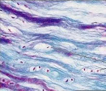
Dense Regular Connective Tissue: Forms ligaments.
Blood Tissue: Present in red marrow for hematopoiesis.
Structure of Bone
Long Bone Anatomy
Diaphysis: Shaft, made of compact bone.
Epiphysis: Ends, mostly spongy bone.
Periosteum: Outer fibrous membrane covering diaphysis.
Endosteum: Lines inner surface of shaft.
Medullary Cavity: Central cavity; contains yellow marrow (fat storage) and red marrow (blood cell formation).
Articular Cartilage: Hyaline cartilage covering epiphyses, reduces friction.
Epiphyseal Plate/Line: Growth plate in young bones; becomes epiphyseal line in adults.
Bone Markings
Projections/Processes: Sites for muscle, tendon, and ligament attachment.
Depressions/Cavities: Passages for nerves and blood vessels.
Microscopic Anatomy of Bone
Osteocytes: Mature bone cells in lacunae.
Lacunae: Small cavities housing osteocytes.
Lamellae: Concentric rings of bone matrix.
Central (Haversian) Canal: Contains blood vessels and nerves.
Osteon (Haversian System): Structural unit of compact bone.
Canaliculi: Tiny canals connecting osteocytes to nutrient supply.
Perforating (Volkmann’s) Canal: Perpendicular canals for blood vessels and nerves.
Bone Formation, Growth, and Remodeling
Ossification
Bone formation (ossification) occurs on hyaline cartilage models or fibrous membranes. Long bone formation involves two major phases:
Osteoblasts cover cartilage model with bone matrix.
Cartilage is replaced by bone matrix, forming medullary cavity.
Growth in Length and Width
Length: Occurs at articular cartilages and epiphyseal plates. New cartilage forms externally; old cartilage is replaced by bone internally.
Width (Appositional Growth): Osteoblasts add bone matrix externally; osteoclasts remove bone internally.
Bone Remodeling
Controlled by calcium ion levels and mechanical stress.
Osteoclasts reabsorb bone; osteoblasts lay down new bone.
Bone Fractures and Repair
Types of Fractures
Closed (Simple) Fracture: Bone breaks but does not penetrate skin.
Open (Compound) Fracture: Bone breaks and penetrates skin.
Fracture Treatment and Healing
Reduction: Realignment of bone (closed or open).
Immobilization: Stabilizes bone for healing (6–8 weeks).
Stages of Bone Repair
Hematoma formation
Fibrocartilage callus formation
Bony callus formation
Bone remodeling
Axial Skeleton
Components
Skull: Cranium (protects brain) and facial bones.
Vertebral Column: Cervical, thoracic, lumbar vertebrae, sacrum, coccyx.
Bony Thorax: Sternum, ribs, thoracic vertebrae.
Appendicular Skeleton
Components
Pectoral Girdle: Clavicle and scapula.
Upper Limbs: Humerus, radius, ulna, carpals, metacarpals, phalanges.
Pelvic Girdle: Ilium, ischium, pubis, sacrum, coccyx.
Lower Limbs: Femur, tibia, fibula, tarsals, metatarsals, phalanges.
Joints
Classification
Functional: Synarthroses (immovable), amphiarthroses (slightly movable), diarthroses (freely movable).
Structural: Fibrous (immobile), cartilaginous (slightly movable), synovial (freely movable).
Synovial Joints
Articular cartilage: Hyaline cartilage on bone ends.
Articular capsule: Fibrous connective tissue enclosing joint.
Joint cavity: Contains synovial fluid.
Reinforcing ligaments: Dense connective tissue.
Bursae: Fluid-filled sacs reducing friction.
Tendon sheath: Elongated bursa wrapping around tendons.
Types of Synovial Joints
Plane: Flat surfaces, gliding movements (e.g., intercarpal joints).
Hinge: Uniaxial movement (e.g., elbow, knee).
Pivot: Rotation around one axis (e.g., atlas/axis).
Condylar: Biaxial movement (e.g., metacarpal/phalangeal joints).
Saddle: Biaxial movement (e.g., thumb).
Ball-and-Socket: Multiaxial movement (e.g., shoulder, hip).
Summary Table: Joint Classes
Functional Class | Structural Class | Mobility | Examples |
|---|---|---|---|
Synarthroses | Fibrous | Immovable | Sutures in skull |
Amphiarthroses | Cartilaginous | Slightly movable | Pubic symphysis, intervertebral joints |
Diarthroses | Synovial | Freely movable | Shoulder, hip, knee |
Additional info: Academic context was added to clarify bone tissue types, joint classifications, and the stages of bone repair. Images included are directly relevant to the explanation of bone structure and tissue types.
