 Indietro
Study Guide: The Skeletal System in Human Anatomy
Guida di studio - Note intelligenti
 Appunti personalizzati basati sui tuoi materiali, ampliati con definizioni chiave, esempi e contesto.
The Skeletal System
Functions of the Skeletal System
The skeletal system is essential for providing structure, protection, and support to the human body. It also plays a critical role in mineral storage, blood cell production, and force transmission.
Shape and Support: The skeleton forms the framework that supports the body and maintains its shape.
Protection: Bones protect vital organs, such as the brain (skull), heart and lungs (rib cage), and spinal cord (vertebral column).
Storage and Production: Bones store minerals (e.g., calcium, phosphorus) and produce red blood cells in the bone marrow.
Force Transmission: The skeleton provides a system for transmitting muscular forces, enabling movement.
Structure of the Skeletal System
The human skeleton consists of approximately 206 bones and over 200 joints, accounting for 12-15% of body weight. It is composed primarily of connective tissue, and bone structure varies to suit specific functions.
Bones: Rigid organs forming the skeleton.
Joints: Articulations between bones, allowing movement.
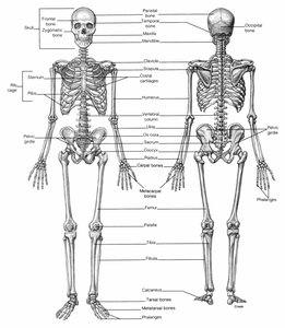
Classification of Bones
Bones are classified by their shapes, which relate to their functions:
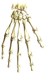
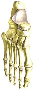
Short Bones: Found in the ankles (tarsals) and wrists (carpals); act as shock absorbers.
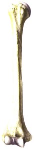
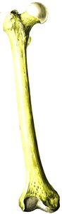
Long Bones: Include the femur and humerus; length greatly exceeds diameter, providing levers for movement.
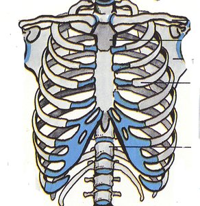
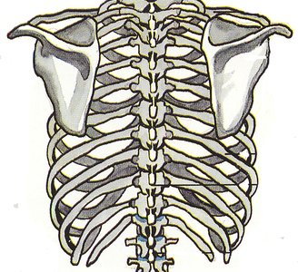
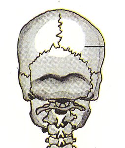
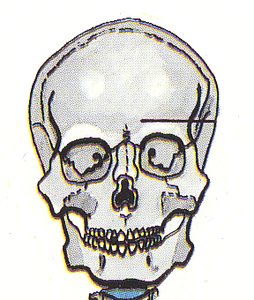
Flat Bones: Skull, scapula, ribs, sternum, and clavicle; protect underlying organs.
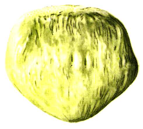
Sesamoid Bones: Patella; embedded within tendons, rounded or oval in shape.
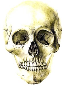
Irregular Bones: Vertebrae and facial bones; fulfill specialized roles.
Axial and Appendicular Skeleton
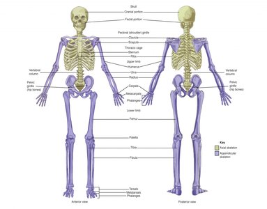
The skeleton is divided into two main regions:
Axial Skeleton: ~80 bones, including the skull, vertebral column, and rib cage. Provides central support and protection.
Appendicular Skeleton: ~126 bones, including the limbs and girdles (pectoral and pelvic). Facilitates movement.
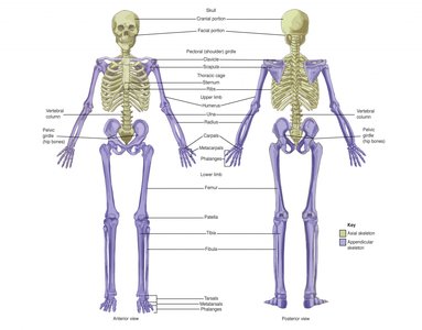
Bone Structure and Examples
Short Bones
Short bones are typically cube-shaped and provide stability and support with limited movement.
Examples: Tarsals (ankle), Carpals (wrist)
Long Bones
Long bones are characterized by a shaft and two ends, and are crucial for movement and support.
Examples: Femur (thigh), Humerus (arm)
Flat Bones
Flat bones are thin, flattened, and often curved, providing protection and broad surfaces for muscle attachment.
Examples: Skull, Scapula, Ribs, Sternum, Clavicle
Sesamoid Bones
Sesamoid bones are small, round bones embedded within tendons, protecting them from stress and wear.
Example: Patella (kneecap)
Irregular Bones
Irregular bones have complex shapes that do not fit into other categories.
Examples: Vertebrae, Facial bones
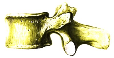
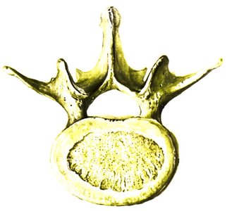
Axial Skeleton Details
Skull
The skull consists of cranial and facial bones, protecting the brain and supporting facial structures.
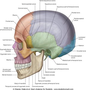
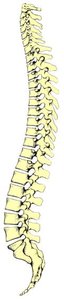
Vertebral Column
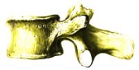
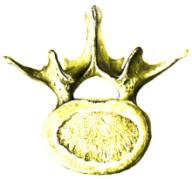
The vertebral column is composed of cervical, thoracic, lumbar, sacral, and coccygeal vertebrae, providing support and flexibility.
7 Cervical Vertebrae: Neck region
12 Thoracic Vertebrae: Chest region
5 Lumbar Vertebrae: Lower back
Sacrum: Mid-line region of buttocks
Coccyx: Tail bone (4-5 fused vertebrae)
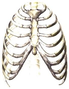
Rib Cage and Sternum
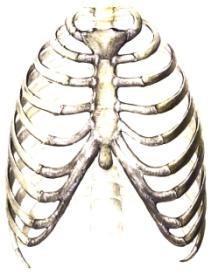
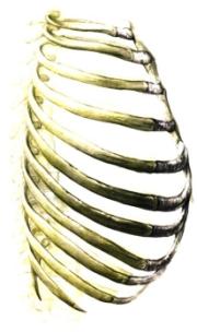
The rib cage consists of twelve pairs of ribs and the sternum, protecting the heart and lungs.
Ribs: Bone and cartilage components
Sternum: Comprised of the manubrium, sternal body, and xiphoid process
Appendicular Skeleton Details
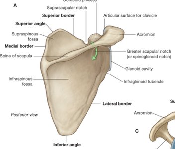
Pectoral Girdle
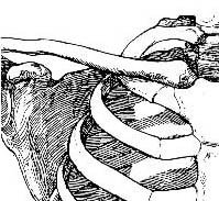
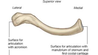
The pectoral girdle consists of the scapula and clavicle, allowing for upper limb mobility.
Sternoclavicular Joint: Only point of attachment between axial skeleton and pectoral girdle
Acromioclavicular Joint: Connects scapula to clavicle
Upper Limb Bones
Humerus: Upper arm bone
Radius and Ulna: Forearm bones
Carpals, Metacarpals, Phalanges: Bones of the wrist and hand
Pelvic Girdle
The pelvic girdle is formed by a pair of os coxae (innominate bones), supporting the bladder and abdominal contents.
Attachments: Posteriorly to sacrum, anteriorly to each other (pubic symphysis), laterally to head of femur
Lower Limb Bones
Femur: Thigh bone
Patella: Sesamoid bone in the knee
Tibia and Fibula: Leg bones
Calcaneus and Talus: Tarsal bones of the ankle
Metatarsals and Phalanges: Bones of the foot
Bone Health and Fitness
Regular physical activity increases bone density and mineralization, while inactivity leads to bone loss. Osteoporosis and osteopenia are disorders characterized by decreased bone mass and density, increasing fracture risk.
Type I Osteoporosis: Post-menopausal, affects 40% of women over 50
Type II Osteoporosis: Age-associated, affects most women and men over 70
Bone Hypertrophy: Increased bone mass due to osteoblast activity
Bone Atrophy: Decreased bone mass due to osteoclast activity
Female Athlete Triad
The Female Athlete Triad is a syndrome involving low energy availability, menstrual disorders, and osteoporosis, commonly seen in adolescent and college-age female athletes.
Low Energy Availability: Often due to eating disorders
Menstrual Disorders: Disrupted menstrual cycles
Osteoporosis: Low bone mineral density
Summary Table: Bone Types and Functions
Bone Type | Example | Function |
|---|---|---|
Short | Tarsals, Carpals | Shock absorption |
Long | Femur, Humerus | Levers for movement |
Flat | Skull, Scapula, Ribs | Protection |
Sesamoid | Patella | Protect tendons |
Irregular | Vertebrae, Facial bones | Specialized roles |
Key Equations
Bone Mass Change:
Additional info: Academic context was added to clarify bone types, functions, and health conditions.
