 Indietro
Study Guide: The Spinal Cord, Spinal Nerves, and Spinal Reflexes
Guida di studio - Note intelligenti
 Appunti personalizzati basati sui tuoi materiali, ampliati con definizioni chiave, esempi e contesto.
Chapter 13: The Spinal Cord, Spinal Nerves, and Spinal Reflexes
The Nervous System: Structural Organization
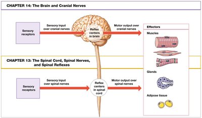
The nervous system is divided into the central nervous system (CNS) and peripheral nervous system (PNS). The CNS includes the brain and spinal cord, which serve as processing centers. The PNS consists of cranial and spinal nerves, responsible for sensory input and motor response pathways. Reflexes are quick, automatic responses triggered by specific stimuli, with spinal reflexes controlled by the spinal cord alone, independent of the brain.
The Spinal Cord: Gross Anatomy
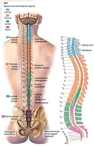
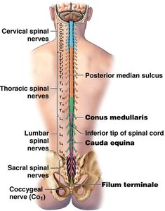
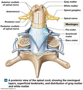
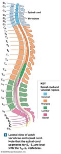
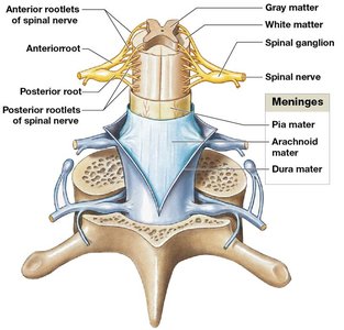
The spinal cord is an organ made of nervous tissue, protected by membranes (meninges) and the vertebral column. It carries sensory and motor information between the brain and most other parts of the body and is the origin of spinal nerves. The spinal cord is about 18 inches (45 cm) long and 0.55 inches (14 mm) wide, extending from the brain to vertebrae L1 and L2. It contains gray matter (neuronal cell bodies) and white matter (myelinated axons), and is divided into four regions: cervical, thoracic, lumbar, and sacral.
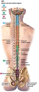
31 segments give rise to spinal nerves.
Bilateral symmetry with grooves dividing left and right sides.
Central canal contains cerebrospinal fluid (CSF).
Enlargements supply nerves to limbs: cervical (shoulder/upper limb) and lumbosacral (pelvis/lower limb).
Spinal Roots and Ganglia
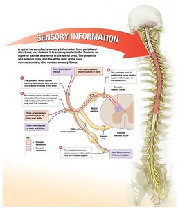
Each spinal nerve attaches to the spinal cord via two roots:
Anterior (ventral) root: axons of motor neurons
Posterior (dorsal) root: axons of sensory neurons
Spinal ganglia (dorsal root ganglia): contain cell bodies of sensory neurons, located between vertebrae
Spinal Nerves: Structure and Branches
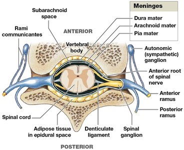
Spinal nerves are formed by the union of anterior and posterior roots and are classified as mixed nerves (containing both sensory and motor fibers). They branch into:
White ramus communicans: myelinated axons to glands and smooth muscle
Gray ramus communicans: unmyelinated fibers to glands and smooth muscle
Posterior ramus: innervates skin/muscles of the back
Anterior ramus: innervates ventrolateral trunk and limbs
Naming Spinal Nerves
Spinal nerves are designated by their vertebral region and number. For example, C1 runs above the first cervical vertebra, C8 is below the seventh cervical vertebra, and others are named for the vertebra above.
Spinal Meninges
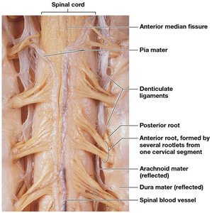
Spinal meninges are specialized membranes surrounding the spinal cord, providing stability, shock absorption, and a pathway for blood vessels. The three layers are:
Dura mater: tough, outer layer
Arachnoid mater: middle, weblike layer
Pia mater: inner, mesh-like layer attached to neural tissue
Meningitis is an infection of the meninges.
Detailed Structure of Meninges
The dura mater contains dense collagen fibers and is continuous with cranial dura mater. The epidural space lies between vertebrae and dura mater, containing loose connective and adipose tissue. The arachnoid mater includes the arachnoid membrane and trabeculae, with the subarachnoid space filled with CSF. The pia mater is firmly attached to neural tissue, with blood vessels on its surface and denticulate ligaments anchoring it to other layers.
Gray Matter and White Matter
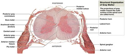
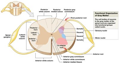
Gray matter consists of neuron cell bodies, neuroglia, and unmyelinated axons, integrating information and initiating commands. It is located deep in the spinal cord and organized into nuclei and horns:
Posterior horns: somatic and visceral sensory nuclei
Anterior horns: somatic motor nuclei
Lateral horns: visceral motor nuclei (thoracic/lumbar segments)
Gray commissure: band around central canal
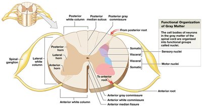
White matter contains myelinated and unmyelinated axons, carrying information. It is organized into three columns:
Posterior white columns: between posterior horns and median sulcus
Anterior white columns: between anterior horns and median fissure
Lateral white columns: on each side between anterior and posterior columns
Tracts are bundles of axons relaying information. Ascending tracts carry sensory information to the brain, while descending tracts carry motor commands to the spinal cord.
Spinal Nerves and Plexuses
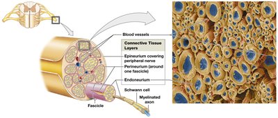
Spinal nerves are surrounded by three connective tissue layers:
Epineurium: outermost, collagen fibers
Perineurium: middle, separates nerve into fascicles
Endoneurium: innermost, surrounds individual axons
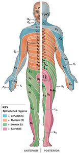
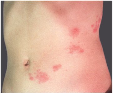
Peripheral nerves are formed as spinal nerves branch and interconnect. Dermatome is a region of skin monitored by a single pair of spinal nerves. Peripheral neuropathies are regional losses of function, often from trauma or compression. Shingles is a viral infection affecting spinal nerves, producing a painful rash along dermatomes.
Nerve Plexuses
A nerve plexus is a complex network of nerve fibers formed from blended fibers of anterior rami of adjacent spinal nerves. This allows multiple spinal nerves to supply the same structures. The four major plexuses are:
Cervical plexus
Brachial plexus
Lumbar plexus
Sacral plexus
Cervical Plexus
Includes anterior rami of C1–C5, innervates scalp, neck, and diaphragm. Major nerves: phrenic (diaphragm), lesser occipital, great auricular, transverse cervical, supraclaviculars, ansa cervicalis.
Brachial Plexus
Anterior rami of C5–T1, innervates pectoral girdle, upper back, and upper limbs. Trunks form cords, which give rise to major nerves: musculocutaneous, radial, median, ulnar.
Lumbar Plexus
Anterior rami of T12–L4, innervates abdominal muscles, thigh, and external genitalia. Major nerves: iliohypogastric, ilio-inguinal, genitofemoral, lateral femoral cutaneous, femoral, obturator.
Sacral Plexus
Anterior rami of L4–S4, innervates gluteal muscles, posterior thigh, leg, perineum. Major nerves: superior/inferior gluteal, posterior femoral cutaneous, sciatic (fibular and tibial branches), pudendal.
Neuronal Pools
Neuronal pools are functional groups of interconnected interneurons, with limited input sources and output destinations. They may stimulate or depress parts of the CNS. Patterns include:
Divergence: spreads information from one neuron to many (common in sensory pathways)
Convergence: several neurons synapse on a single neuron (e.g., control of diaphragm)
Serial processing: information moves sequentially through neurons (e.g., pain signals)
Parallel processing: several neurons process the same information simultaneously (e.g., stepping on a sharp object)
Reverberation: positive feedback loop maintains activity (e.g., breathing, muscle coordination)
Reflexes and Reflex Arcs
A neural reflex is a rapid, automatic response to specific stimuli, helping preserve homeostasis. Reflexes follow a reflex arc:
Stimulus activates a receptor
Activation of a sensory neuron
Information processing in CNS (interneurons)
Activation of a motor neuron
Response by a peripheral effector (muscle or gland)
Reflexes are classified by development (innate/acquired), motor response (somatic/visceral), complexity (monosynaptic/polysynaptic), and site of processing (spinal/cranial).
Spinal Reflexes
Monosynaptic spinal reflexes (e.g., stretch reflex) regulate skeletal muscle length and are very rapid. The muscle spindle is the sensory receptor involved, made of intrafusal fibers surrounded by extrafusal fibers. Sensory neurons detect stretch and synapse with motor neurons, stimulating muscle contraction to prevent overstretching. Gamma efferents adjust spindle sensitivity during voluntary contraction.
Postural Reflexes
Help maintain upright posture, involve stretch reflexes and complex polysynaptic reflexes, and require constant fine adjustments.
Polysynaptic Reflexes
Allow for more complex responses, involving interneurons that control multiple muscle groups. Examples include tendon reflex (prevents excessive tension), withdrawal reflexes (move body part from pain), and crossed-extensor reflexes (coordinate limbs during injury).
Reciprocal Inhibition
For flexor reflexes to work, antagonistic muscles must be inhibited by interneurons. When flexors contract, extensors relax, and vice versa.
Reflex Arcs
Reflex arcs can be ipsilateral (same side as stimulus) or contralateral (opposite side, as in crossed-extensor reflexes).
Characteristics of Polysynaptic Reflexes
Involve pools of interneurons (excitation/inhibition)
Involve more than one spinal segment
Involve reciprocal inhibition
Have reverberating circuits
Several reflexes may cooperate for coordinated response
Brain Influence on Spinal Reflexes
Although spinal reflexes are automatic, the brain can facilitate or inhibit them. Voluntary movements can activate the same motor patterns as reflexes. Higher centers can reinforce reflexes (general facilitation) or inhibit them (via inhibitory neurons and IPSPs). The plantar reflex (toe-curling) is normal in adults, while the Babinski reflex (toe fanning) is normal in infants but may indicate CNS damage in adults.
Key Terms and Concepts
Reflex arc: pathway followed by nerve impulses to produce a reflex
Monosynaptic reflex: single synapse, fast response
Polysynaptic reflex: multiple synapses, slower, more complex response
Dermatome: region of skin supplied by a single spinal nerve
Muscle spindle: sensory receptor for stretch reflex
Golgi tendon organ: sensory receptor for tendon reflex
Summary Table: Spinal Cord Regions and Functions
Region | Function | Major Nerves |
|---|---|---|
Cervical | Neck, diaphragm, upper limbs | Phrenic, lesser occipital |
Brachial | Pectoral girdle, upper limbs | Musculocutaneous, radial, median, ulnar |
Lumbar | Abdominal muscles, thigh, genitalia | Femoral, obturator |
Sacral | Gluteal muscles, posterior thigh, leg, perineum | Sciatic, pudendal |
Example: The patellar reflex is a monosynaptic stretch reflex that helps maintain posture by causing contraction of the quadriceps muscle when the patellar tendon is tapped.
Additional info: The notes above expand on brief points with academic context, definitions, and examples to ensure completeness and clarity for exam preparation.
