 Indietro
CH 13: The Spinal Cord, Spinal Nerves, and Somatic Reflexes
Guida di studio - Note intelligenti
 Appunti personalizzati basati sui tuoi materiali, ampliati con definizioni chiave, esempi e contesto.
The Spinal Cord
Functions of the Spinal Cord
The spinal cord is a vital structure in the central nervous system, responsible for transmitting information between the brain and the rest of the body. It performs several key functions:
Conduction: Nerve fibers conduct sensory and motor information up and down the spinal cord.
Neural Integration: Spinal neurons receive input from multiple sources, integrate it, and execute appropriate output (e.g., bladder control).
Locomotion: Central pattern generators in the spinal cord coordinate repetitive muscle contractions for walking.
Reflexes: Involuntary responses to stimuli, essential for posture, coordination, and protection.
Surface Anatomy of the Spinal Cord
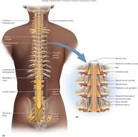
The spinal cord is a cylindrical structure arising from the brainstem at the foramen magnum. It occupies the upper two-thirds of the vertebral canal and ends at the level of L1 or slightly beyond. The cord gives rise to 31 pairs of spinal nerves, each supplying a segment of the body.
Regions: Cervical, thoracic, lumbar, and sacral.
Cervical enlargement: Supplies nerves to the upper limbs.
Lumbosacral enlargement: Supplies nerves to the pelvic region and lower limbs.
Medullary cone (conus medullaris): Inferior point of the cord.
Cauda equina: Bundle of nerve roots from L2 to S5, resembling a horse's tail.
Meninges of the Spinal Cord
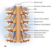
The spinal cord is protected by three fibrous membranes called meninges:
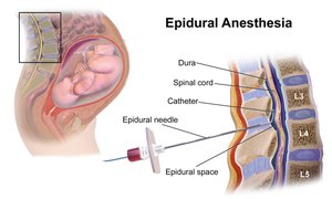
Dura mater: Tough, thick membrane composed of dense irregular connective tissue. The epidural space between the dura and vertebral bones contains blood vessels, adipose tissue, and loose connective tissue. Anesthetics can be introduced here (epidural anesthesia).
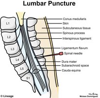
Arachnoid mater: Adheres to the dura and is separated from the pia mater by the subarachnoid space, which is filled with cerebrospinal fluid (CSF). The lumbar cistern, located inferior to the medullary cone, is the site for lumbar puncture (spinal tap).
Pia mater: Delicate membrane that follows the contours of the spinal cord and forms the terminal filum, which anchors the cord.
Cross-Sectional Anatomy of the Spinal Cord
The spinal cord consists of gray matter and white matter:
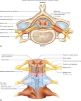
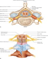
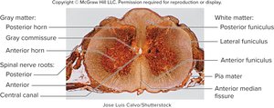
Gray matter: Contains neuron cell bodies, dendrites, and axons; shaped like a butterfly in cross-section. Includes posterior (dorsal) horns (sensory), anterior (ventral) horns (motor), and lateral horns (sympathetic nervous system).
White matter: Composed of myelinated axons; organized into posterior, lateral, and anterior funiculi (columns), each subdivided into tracts.
Spinal Tracts
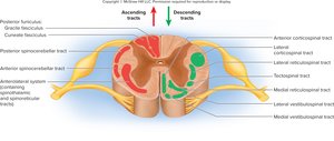
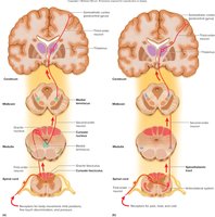
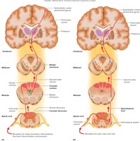
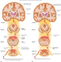
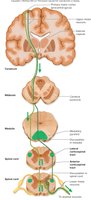
Spinal tracts are bundles of axons with similar origin, destination, and function. They are classified as ascending (sensory) or descending (motor):
Ascending tracts: Carry sensory information to the brain. Pathways typically involve three neurons: first-order (detects stimulus), second-order (to thalamus), and third-order (to cerebral cortex).
Descending tracts: Carry motor commands from the brain to effectors. Pathways involve two neurons: upper motor neuron (originates in cortex/brainstem) and lower motor neuron (in spinal cord/brainstem).
Decussation: Many tracts cross the midline, resulting in contralateral control.
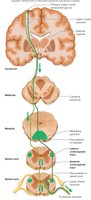
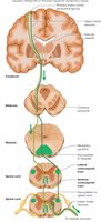
Clinical Correlation: Poliomyelitis and Amyotrophic Lateral Sclerosis (ALS)
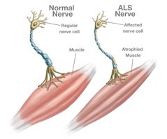
Both diseases cause destruction of motor neurons, leading to skeletal muscle atrophy:
ALS: Progressive muscular weakness, difficulty speaking/swallowing, and atrophy. Sensory and intellectual functions remain unaffected.
The Spinal Nerves
Anatomy of Nerves and Ganglia
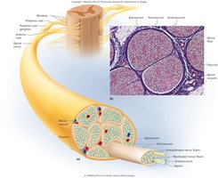
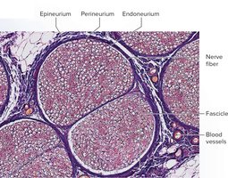
Spinal nerves communicate between the spinal cord and the body. A nerve is a cord-like organ composed of numerous nerve fibers (axons) bound by connective tissue:
Schwann cells: Form neurilemma and myelin sheath around axons.
Endoneurium: Surrounds individual axons.
Perineurium: Wraps fascicles (bundles of nerve fibers).
Epineurium: Wraps the entire nerve.
Blood vessels: Penetrate coverings, providing blood supply.
Classification of Nerves
Sensory nerves: Carry signals from receptors to CNS (rare).
Motor nerves: Carry signals from CNS to effectors.
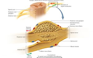
Mixed nerves: Contain both sensory and motor fibers (most common).
Ganglion: Cluster of nerve cell bodies outside CNS, enveloped in epineurium.
Spinal Nerve Structure
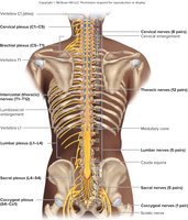
31 pairs: 8 cervical, 12 thoracic, 5 lumbar, 5 sacral, 1 coccygeal.
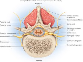
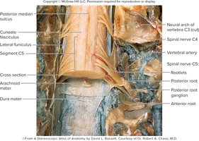
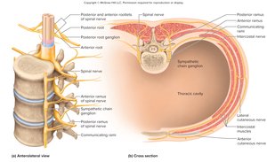
Proximal branches: Posterior (dorsal) root (sensory), anterior (ventral) root (motor).
Posterior root ganglion: Contains sensory neuron cell bodies.
Cauda equina: Roots from L2 to Co1.
Distal Branches and Rami
Anterior ramus: Forms intercostal nerves (thoracic) or nerve plexuses (other regions).
Posterior ramus: Innervates muscles, joints, and skin of the back.
Clinical Correlation: Shingles
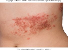
Varicella-zoster virus: Remains dormant in posterior root ganglia after chickenpox.
Shingles: Reactivation causes painful skin lesions along the path of affected nerve.
Nerve Plexuses
Major Nerve Plexuses
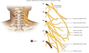
Cervical plexus (C1-C5): Supplies neck and phrenic nerve to diaphragm.
Brachial plexus (C5-T1): Supplies upper limb, shoulder, and neck.
Lumbar plexus (L1-L4): Supplies abdominal wall, anterior thigh, and genitalia.
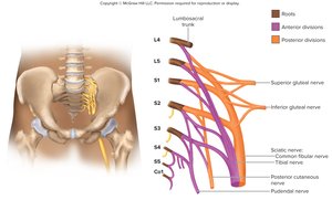
Sacral plexus (L4-S4): Supplies lower trunk and limb, including sciatic nerve.
Coccygeal plexus (S4-Co1): Supplies coccygeal region.
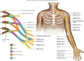
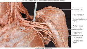
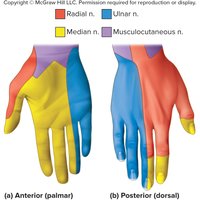
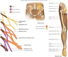
Nerve Injuries
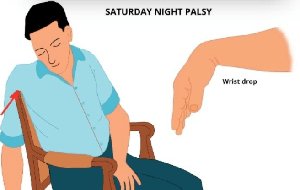
Radial nerve injury: May cause wrist drop due to paralysis of extensor muscles.
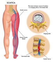
Sciatic nerve injury: Sciatica causes sharp pain along the posterior leg, often due to herniated disc.
Dermatomes and Cutaneous Innervation
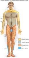
Dermatome Map
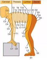
A dermatome is a specific area of skin that conveys sensory input to a spinal nerve. Dermatome maps show the cutaneous regions innervated by each spinal nerve. Overlap between dermatomes means that three successive nerves must be anesthetized for complete loss of sensation in one area.
Somatic Reflexes
The Nature of Reflexes
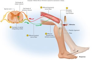
Reflexes are quick, involuntary, stereotyped reactions of glands or muscles to stimulation. Somatic reflexes involve skeletal muscle, while visceral reflexes involve internal organs.
Reflex arc: Pathway includes somatic receptors, afferent nerve fibers, integrating center, efferent nerve fibers, and effectors.
The Muscle Spindle
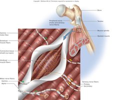
Muscle spindles are stretch receptors embedded in skeletal muscles, serving as proprioceptors to monitor position and movement. They inform the brain of muscle length and enable coordinated movement and posture.
The Stretch Reflex
The stretch (myotatic) reflex occurs when a muscle is stretched and contracts to maintain equilibrium and posture. The patellar (knee-jerk) reflex is a classic example, mediated by a monosynaptic reflex arc.
Reciprocal inhibition: Prevents muscles from working against each other by inhibiting antagonists when agonists are excited.
The Flexor (Withdrawal) Reflex
The flexor reflex is a quick contraction of flexor muscles resulting in withdrawal from an injurious stimulus. It involves a polysynaptic reflex arc.
The Crossed Extension Reflex
The crossed extension reflex contracts extensor muscles in the limb opposite to the one withdrawn, maintaining balance. The flexor reflex uses an ipsilateral arc, while the crossed extension reflex uses a contralateral arc.
The Tendon Reflex
Tendon organs are proprioceptors in tendons near their junction with muscle. The tendon reflex responds to excessive tension, inhibiting muscle contraction to prevent injury.
Spinal Cord Trauma
Types of Paralysis
Complete transection: Severance of the cord results in loss of motor control below the injury.
Paraplegia: Paralysis of both lower limbs.
Quadriplegia: Paralysis of all four limbs.
Hemiplegia: Paralysis on one side of the body.
Paresis: Partial paralysis or weakness.
Key Terms and Concepts
Proprioception: Nonvisual sense of body position and movement.
Decussation: Crossing of nerve fibers from one side of the CNS to the other.
Contralateral: Origin and destination on opposite sides.
Ipsilateral: Origin and destination on the same side.
Sample Table: Spinal Cord Regions and Functions
Region | Function |
|---|---|
Cervical | Upper limb innervation |
Thoracic | Trunk innervation |
Lumbar | Lower limb innervation |
Sacral | Pelvic region and lower limb innervation |
Sample Equation: Nerve Signal Pathway
For ascending tracts:
For descending tracts:
