 Indietro
Study Guide: The Urinary System (Anatomy & Physiology)
Guida di studio - Note intelligenti
 Appunti personalizzati basati sui tuoi materiali, ampliati con definizioni chiave, esempi e contesto.
The Urinary System: Overview
Functions of the Urinary System
The urinary system is essential for maintaining the body's internal environment by regulating water, solute concentrations, and removing metabolic wastes. It also plays roles in hormone production and vitamin D activation.
Regulation of water and solute concentration: Maintains total body water volume and solute concentration.
Ion regulation: Controls concentrations of ions in the extracellular fluid (ECF).
Acid-base balance: Ensures long-term acid-base balance.
Excretion: Removes metabolic wastes, toxins, and drugs.
Hormone production: Produces erythropoietin (stimulates RBC production) and renin (regulates blood pressure).
Vitamin D activation: Converts vitamin D to its active form.
Gluconeogenesis: Generates glucose during prolonged fasting.
Organs of the Urinary System
The urinary system consists of four main organs, each with a distinct function.
Kidneys: Filter blood and produce urine.
Ureters: Transport urine from kidneys to bladder.
Urinary Bladder: Temporarily stores urine.
Urethra: Carries urine out of the body.
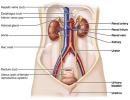
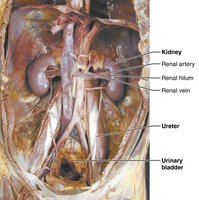
Kidney Anatomy
Location and External Anatomy
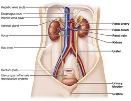
The kidneys are retroperitoneal organs located in the superior lumbar region, protected by the rib cage. The right kidney sits lower due to the liver.
Renal hilum: Medial cleft where vessels, nerves, and ureters enter/exit.
Adrenal glands: Rest atop each kidney.
Supportive tissue: Includes renal fascia, perirenal fat capsule, and fibrous capsule.
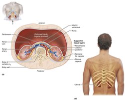
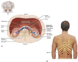
Internal Gross Anatomy
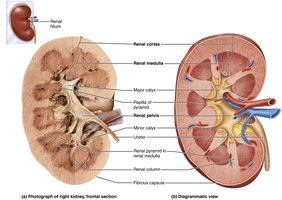
The kidney has three distinct regions: cortex, medulla, and pelvis. Each region has specialized structures for urine formation and transport.
Renal cortex: Superficial, granular region.
Renal medulla: Deep region with cone-shaped pyramids; pyramids are separated by renal columns.
Renal pelvis: Funnel-shaped tube continuous with the ureter; branches into major and minor calyces.
Urine Flow Through the Kidney
Urine is formed in the medullary pyramids and follows a specific pathway before exiting the body.
Pathway: Papillae → Minor calyx → Major calyx → Renal pelvis → Ureter → Urinary bladder → Urethra
Renal Blood Supply
Oxygenated Blood Vessels
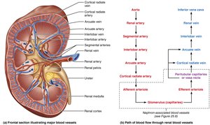
The kidneys receive about 25% of cardiac output per minute. Blood enters via the renal arteries and passes through several branches before reaching the glomerulus.
Pathway: Aorta → Renal artery → Segmental artery → Interlobar artery → Arcuate artery → Cortical radiate artery → Afferent arteriole → Glomerulus
Deoxygenated Blood Vessels
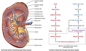
Blood exits the kidney via the renal vein, following a pathway through several venous branches.
Pathway: Cortical radiate vein → Arcuate vein → Interlobar vein → Renal vein → Inferior vena cava
Nephrons: The Functional Unit
Structure and Types
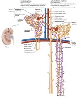
Each kidney contains about 1 million nephrons, which are responsible for filtering blood and forming urine. Nephrons are located in both the cortex and medulla.
Renal corpuscle: Includes the glomerulus and glomerular (Bowman's) capsule.
Renal tubule: Consists of the proximal convoluted tubule (PCT), nephron loop (Loop of Henle), and distal convoluted tubule (DCT).
Collecting ducts: Receive filtrate from multiple nephrons.
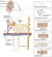
Cortical vs. Juxtamedullary Nephrons
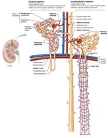
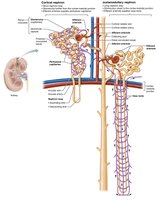
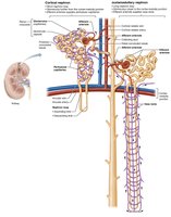
Cortical nephrons: 85% of nephrons; located mostly in the cortex with short loops.
Juxtamedullary nephrons: Originate near the cortex-medulla junction; have long loops extending deep into the medulla.
Nephron Capillary Beds
Nephrons are associated with three capillary beds: glomerulus, peritubular capillaries, and vasa recta.
Glomerulus: Fenestrated capillaries for filtration.
Peritubular capillaries: Surround renal tubules; absorb solutes and water.
Vasa recta: Long, straight vessels in juxtamedullary nephrons; important for urine concentration.
Juxtaglomerular Complex (JGC)
Structure and Function
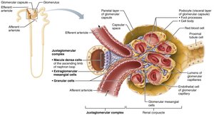
The JGC is a specialized region where the ascending limb of the nephron loop contacts the afferent arteriole. It regulates blood pressure and filtration rate.
Macula densa: Chemoreceptors monitoring NaCl concentration.
Extraglomerular mesangial cells: Pass regulatory signals.
Granular (JG) cells: Mechanoreceptors monitoring blood pressure; secrete renin.
Urine Formation: Major Renal Processes
Three Steps of Urine Formation
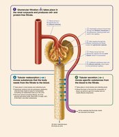
Urine formation involves three main processes: glomerular filtration, tubular reabsorption, and tubular secretion.
Glomerular filtration: Passive process where hydrostatic pressure forces fluids and solutes through the filtration membrane.
Tubular reabsorption: Reclaims most tubule contents and returns them to the blood.
Tubular secretion: Moves selected substances from blood into the filtrate for excretion.
Glomerular Filtration
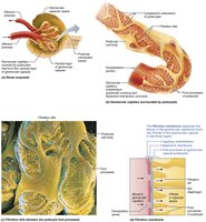
The filtration membrane consists of three layers: fenestrated endothelium, basement membrane, and podocyte foot processes. It allows passage of water and small solutes, but blocks blood cells and most proteins.
Filtration membrane: Permits water, glucose, amino acids, and nitrogenous wastes; plasma proteins remain in blood.
Pressures: Outward hydrostatic pressure promotes filtration; inward hydrostatic and colloid osmotic pressures inhibit filtration.
Net Filtration Pressure (NFP):
Glomerular Filtration Rate (GFR): Volume of filtrate formed per minute (~125 mL/min); directly proportional to NFP, surface area, and membrane permeability.
Regulation of GFR
GFR is tightly regulated to maintain homeostasis and blood pressure. Intrinsic controls (renal autoregulation) operate when MAP is 80–180 mmHg; extrinsic controls (nervous/endocrine) override when MAP is outside this range.
Myogenic response: Smooth muscle contracts/relaxes to regulate blood flow.
Tubuloglomerular feedback: Macula densa cells respond to NaCl levels, adjusting afferent arteriole diameter.
Tubular Reabsorption
Most filtrate is reabsorbed into the blood, especially organic nutrients. Water and ions are regulated by hormones.
Reabsorption sites: PCT, nephron loop, DCT, collecting duct.
Regulated substances: Water, Ca2+, Mg2+, K+, Na+.
Tubular Secretion
Tubular secretion moves substances from blood into the filtrate, aiding in pH control and waste removal.
Functions: Controls blood pH, disposes of drugs/metabolites, secretes excess K+.
Sites: PCT, ascending nephron loop, DCT, collecting duct.
Assessing Renal Function and Urinalysis
Renal Function Tests
Renal function is assessed by analyzing blood and urine for nitrogenous wastes. Renal clearance tests measure GFR.
Monitored substances: Urea, blood urea nitrogen (BUN), uric acid, creatinine.
Renal clearance: Volume of plasma cleared of a substance per unit time.
Urinalysis
Urinalysis evaluates urine's chemical, macroscopic, and microscopic properties to diagnose disease or infection.
Chemical composition: 95% water, 5% solutes (urea, uric acid, creatinine, ions).
Microscopic components: Plasma proteins, RBCs, WBCs, bile pigments.
Physical characteristics: Color (clear, pale to deep yellow), odor (aromatic), pH (4.5–8.0), specific gravity (1.001–1.035).
Ureters, Urinary Bladder, and Urethra
Ureters
Ureters are slender tubes that convey urine from kidneys to bladder via peristalsis. They are retroperitoneal and attach obliquely to the bladder wall, preventing backflow.
Urinary Bladder
The bladder is a muscular sac for temporary urine storage. Its location differs between males and females.
Openings: Ureters and urethra; trigone area is prone to infection.
Wall layers: Mucosa (transitional epithelium), muscular layer (detrusor), fibrous adventitia.
Capacity: Moderately full bladder holds ~500 mL; can expand to 1,000 mL.
Urethra
The urethra is a thin-walled tube that transports urine out of the body. It has two sphincters: internal (involuntary, smooth muscle) and external (voluntary, skeletal muscle).
Male vs. Female Urethra
Male urethra: Three segments; carries both urine and semen.
Female urethra: Shorter; carries only urine.
