 Indietro
Study Notes: Nervous System Structure and Function
Guida di studio - Note intelligenti
 Appunti personalizzati basati sui tuoi materiali, ampliati con definizioni chiave, esempi e contesto.
Nervous System Overview
Functions of the Nervous System
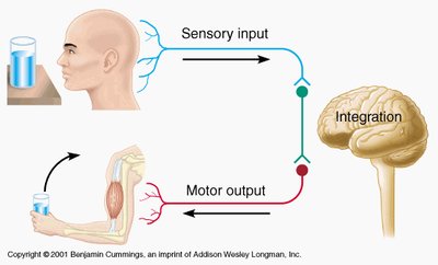
The nervous system is the body's primary control center and communications network. It is responsible for collecting sensory information, integrating and processing that information, and initiating motor responses. These functions are categorized as:
Sensory Function: Collects information from internal and external environments via sensory receptors.
Integrative Function: Processes and evaluates sensory input, allowing for decision-making and response planning.
Motor Function: Responds to information by activating muscles or glands.
Organization of the Nervous System
Structural Divisions
The nervous system is divided into two main structural components:
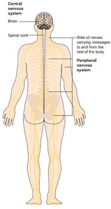
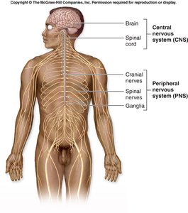
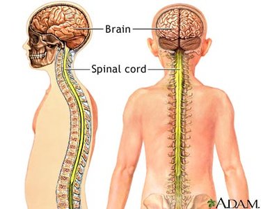
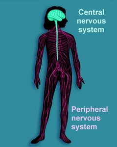
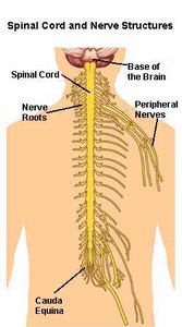
Central Nervous System (CNS): Consists of the brain and spinal cord. It is responsible for processing and integrating information.
Peripheral Nervous System (PNS): Includes all nerves outside the CNS, such as cranial nerves, spinal nerves, and ganglia. It connects the CNS to the rest of the body.
Functional Divisions
The nervous system is also divided functionally:
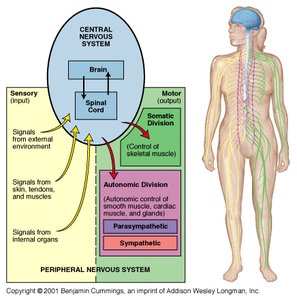
Sensory (Afferent) Division: Brings sensory information from receptors to the CNS.
Motor (Efferent) Division: Carries motor commands from the CNS to effectors (muscles and glands).
Peripheral Nervous System Details
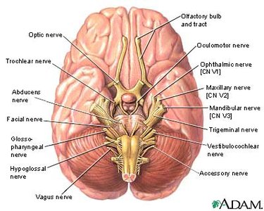
Cranial Nerves: 12 pairs, primarily serving the head and neck.
Spinal Nerves: 31 pairs, serving the rest of the body.
Ganglia: Groups of neuron cell bodies in the PNS.
Functional Divisions of the PNS
Sensory (Afferent) Division
The sensory division transmits information from sensory receptors to the CNS. It includes:
Somatic Sensory Receptors: Monitor external conditions and body position.
Visceral Sensory Receptors: Monitor internal conditions and organ system status.
Motor (Efferent) Division
The motor division transmits commands from the CNS to effectors. It is subdivided into:
Somatic Nervous System: Controls voluntary movements via skeletal muscles.
Autonomic Nervous System: Controls involuntary functions via smooth muscle, cardiac muscle, and glands. It has two major divisions:
Sympathetic Division: Prepares the body for 'fight or flight' responses.
Parasympathetic Division: Promotes 'rest and digest' activities.
Nervous Tissue
Cell Types
Nervous tissue consists of two main cell types:
Neurons: The basic structural and functional units of the nervous system. They conduct electrical impulses and are specialized for communication.
Neuroglial Cells (Glia): Support, protect, and nourish neurons. They can multiply and divide, and are involved in repair and defense.
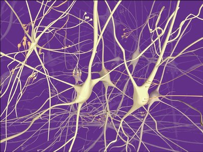
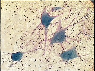
Structure of a Typical Neuron
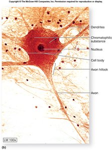
Neurons have specialized structures for signal reception and transmission:
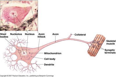
Cell Body (Soma): Contains the nucleus and organelles.
Dendrites: Receive signals and transmit them toward the cell body.
Axon: Propagates signals away from the cell body. May be covered by a myelin sheath for faster conduction.
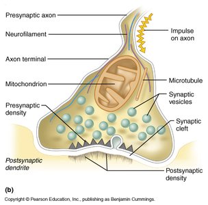
Axon Terminals (Synaptic Knobs): Release neurotransmitters to communicate with other cells.
Neuron Classification
Functional Classes
Afferent (Sensory) Neurons: Carry signals toward the CNS from sensory receptors.
Efferent (Motor) Neurons: Carry signals away from the CNS to effector organs.
Interneurons (Association Neurons): Located entirely within the CNS, connecting afferent and efferent neurons.
Structural Classes
Bipolar Neurons: Found in sensory organs like the retina and olfactory epithelium.
Pseudounipolar Neurons: Sensory neurons with a single process that splits into two branches.
Multipolar Neurons: Motor neurons and interneurons with multiple dendrites and a single axon.
Myelin Sheath and Saltatory Conduction
Myelin Sheath
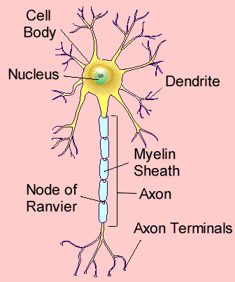
The myelin sheath is a lipid-rich covering that insulates axons and increases the speed of electrical impulse conduction. It is formed by:
Oligodendrocytes: In the CNS.
Schwann Cells (Neurolemmocytes): In the PNS.
Saltatory Conduction
Saltatory conduction is the process by which action potentials jump from one node of Ranvier to the next, greatly increasing conduction speed compared to unmyelinated axons.
Neuroglia (Glial Cells)
Types and Functions
Astrocytes: Support neurons, form the blood-brain barrier, repair injuries, and regulate neurotransmitter activity.
Oligodendrocytes: Produce myelin in the CNS.
Schwann Cells: Produce myelin in the PNS.
Ependymal Cells: Line CNS cavities and circulate cerebrospinal fluid.
Microglia: Phagocytic cells that defend against pathogens.
Satellite Cells: Support neuron cell bodies in the PNS.
Synapses
Structure and Function
A synapse is a functional connection between two neurons, allowing for communication via electrical or chemical signals. The main components include:
Presynaptic Cell: Sends the signal.
Postsynaptic Cell: Receives the signal.
Synaptic Cleft: The gap between the two cells.
Synaptic Vesicles: Contain neurotransmitters released into the cleft.
Central Nervous System: Brain and Spinal Cord
Gray Matter and White Matter
Gray Matter: Contains cell bodies, dendrites, unmyelinated axons, axon terminals, and neuroglia.
White Matter: Composed of myelinated axons.
Brain Divisions
Brainstem: Medulla oblongata, pons, midbrain.
Diencephalon: Thalamus, hypothalamus, pineal body.
Cerebellum: Coordinates movement and balance.
Cerebrum: Two hemispheres, each with six lobes (frontal, parietal, occipital, temporal, insula, limbic).
Spinal Cord
Structure and Function
The spinal cord is a reflex center and conduction pathway, extending from the foramen magnum to L1 or L2. It contains:
Gray Matter: Central bat-shaped area for processing.
White Matter: Columns for relay of motor and sensory tracts.
Spinal Nerves: 31 pairs attached to the cord.
Reflex Arcs
Components of a Reflex Arc
Receptor: Detects stimulus.
Afferent Pathway: Carries signal to CNS.
Integrating Center: Processes input in CNS.
Efferent Pathway: Carries response to effector.
Effector: Executes response.
Example: Patellar Reflex
Stretch Receptor: Muscle spindle.
Stimulus: Tap on patellar ligament.
Effector: Quadriceps femoris muscle.
Action: Contraction extends knee joint.
Summary Table: Structural and Functional Divisions of the Nervous System
Division | Components | Function |
|---|---|---|
Central Nervous System (CNS) | Brain, Spinal Cord | Integration, processing, control |
Peripheral Nervous System (PNS) | Cranial nerves, Spinal nerves, Ganglia | Communication between CNS and body |
Sensory (Afferent) Division | Somatic & Visceral Sensory Receptors | Transmit sensory information to CNS |
Motor (Efferent) Division | Somatic & Autonomic Nervous Systems | Transmit motor commands from CNS |
Additional info: Expanded explanations and examples were added for clarity and completeness.
