 Indietro
Study Notes: The Endocrine System (Anatomy & Physiology, Chapter 16)
Guida di studio - Note intelligenti
 Appunti personalizzati basati sui tuoi materiali, ampliati con definizioni chiave, esempi e contesto.
The Endocrine System
Overview of the Endocrine System
The endocrine system is one of the two major regulatory systems of the body, alongside the nervous system. It consists of organs that synthesize and secrete chemical messengers called hormones into the blood, which interact with specific target cells possessing receptors for those hormones.
Hormones: Chemical messengers secreted into the blood to regulate various physiological processes.
Target Cells: Cells with specific receptors for hormones; tissues containing these cells are called target tissues.
Receptors: Proteins on or within target cells that bind hormones and trigger cellular changes.
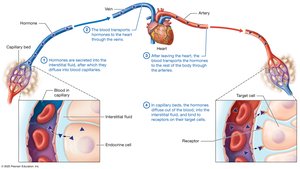
Comparison of the Endocrine and Nervous Systems
The endocrine system differs from the nervous system in its method of communication and speed of response.
Endocrine System: Hormones are secreted into interstitial fluid, diffuse into blood capillaries, and are transported throughout the body. Effects are slower but longer-lasting.
Nervous System: Uses direct synaptic connections for rapid, short-lived responses.
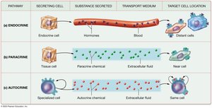
Paracrine and Autocrine Signals
Not all chemical messengers act as classic hormones.
Classic Endocrine Signals: Hormones secreted into the blood to affect distant targets.
Paracrine Signals: Chemicals secreted into extracellular fluid to influence nearby cells.
Autocrine Signals: Chemicals secreted by a cell to elicit effects from the same cell.
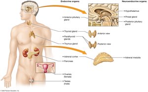
Overview of Endocrine Organs
Endocrine glands are ductless organs that secrete hormones into the interstitial fluid for transport by the bloodstream.
Primary Endocrine Organs: Anterior pituitary, thyroid, parathyroid, adrenal cortices, pancreas, thymus, ovaries/testes.
Secondary Endocrine Glands: Heart, kidneys, small intestine, adipose tissue.
Neuroendocrine Organs: Hypothalamus, pineal gland, adrenal medulla (nervous tissue that secretes hormones).
Classes of Hormones
Hormones are classified based on their chemical structure and solubility.
Amino Acid-Based Hormones: Derived from amino acids; mostly hydrophilic.
Peptide/Protein Hormones: Chains of amino acids; generally hydrophilic.
Steroid Hormones: Derived from cholesterol; hydrophobic and lipid-soluble.
Hormone Transport in Blood
Free Hormones: Small, hydrophilic hormones travel freely in plasma.
Bound Hormones: Hydrophobic hormones form complexes with binding proteins, extending their lifespan and providing a reservoir.
Target Cells and Receptors
Specificity: Receptors are highly specific for their hormones and can bind at very low concentrations.
Multiple Receptors: Some hormones bind to multiple cell types or receptors, producing different effects.
Location: Receptors may be embedded in the plasma membrane (for hydrophilic hormones) or in the cytosol/nucleus (for hydrophobic hormones).
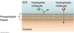
Mechanisms of Hormone Action
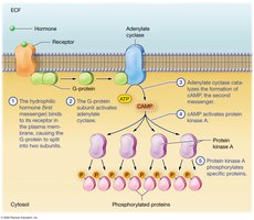
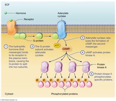
Hydrophilic Hormones: Bind to cell surface receptors and often use second-messenger systems (e.g., cAMP pathway).
Second Messenger System: Hormone binding activates G-proteins, which activate adenylate cyclase, forming cAMP from ATP. cAMP activates protein kinase A, which phosphorylates proteins, amplifying the signal.
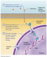
Hydrophobic Hormones: Diffuse into target cells, bind to intracellular receptors, and interact with DNA at hormone-response elements to regulate gene expression.
Regulation of Hormone Secretion
Hormone secretion is regulated by various stimuli and feedback mechanisms.
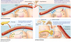
Hormonal Stimuli: Endocrine cells respond to other hormones (e.g., hypothalamic releasing hormones).
Humoral Stimuli: Endocrine cells respond to changes in ion or compound concentrations in blood (e.g., insulin release in response to glucose).
Neural Stimuli: Endocrine cells respond to nervous system signals (e.g., adrenal medulla secretion).
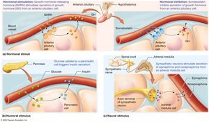
Negative Feedback Regulation
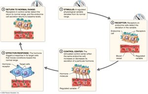
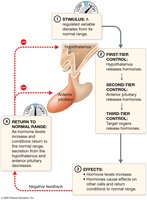
Most hormone secretion is regulated by negative feedback loops.
Stimulus: Physiological variable deviates from normal range.
Receptor: Endocrine cell receptors detect deviation.
Control Center: Endocrine cell increases or decreases hormone secretion.
Effector/Response: Hormone triggers response in target cells, restoring normal range.
Return to Normal: Secretion returns to baseline.
Structure of the Hypothalamus and Pituitary Gland
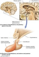
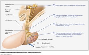
The hypothalamus connects to the pituitary gland via the infundibulum.
Anterior Pituitary (Adenohypophysis): True gland composed of hormone-secreting cells.
Posterior Pituitary (Neurohypophysis): Nervous tissue that stores neurohormones.
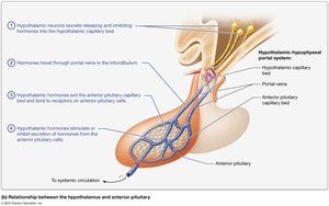
Hypothalamic-Hypophyseal Portal System: Capillaries and portal veins connect hypothalamus and anterior pituitary.
Hormones of the Hypothalamus and Posterior Pituitary
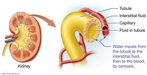
Antidiuretic Hormone (ADH): Water retention; increases water reabsorption in kidneys by inserting aquaporins.
Oxytocin: Stimulates uterine contractions and milk ejection; operates via positive feedback.
Functional Relationship of the Hypothalamus and Anterior Pituitary
The hypothalamus produces releasing and inhibiting hormones that control the anterior pituitary, which in turn secretes tropic hormones affecting other glands.
Thyroid-Stimulating Hormone (TSH): Stimulates thyroid gland.
Adrenocorticotropic Hormone (ACTH): Stimulates adrenal cortex.
Prolactin: Stimulates mammary gland growth and milk production.
Luteinizing Hormone (LH) & Follicle-Stimulating Hormone (FSH): Control gonadal function.
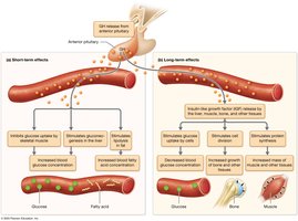
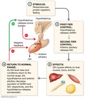
Growth Hormone (GH)
Short-Term Effects: Promotes fat breakdown, gluconeogenesis, and inhibits glucose uptake.
Long-Term Effects: Stimulates IGF production, protein synthesis, cell division, bone and muscle growth.
Structure of the Thyroid and Parathyroid Glands
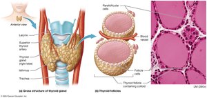
Thyroid Gland: Located in the anterior neck; composed of thyroid follicles filled with colloid.
Parafollicular Cells: Produce calcitonin.
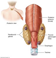
Parathyroid Glands: Usually four glands on the posterior thyroid; chief cells produce parathyroid hormone.
Thyroid Hormones: Metabolic Regulators
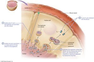
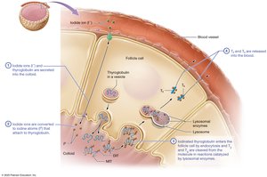
Triiodothyronine (T3): Three iodine atoms; more active.
Thyroxine (T4): Four iodine atoms; converted to T3 in target cells.
Effects: Regulate basal metabolic rate, thermoregulation, growth, and development; synergize with sympathetic nervous system.
Regulation of Thyroid Hormone Production
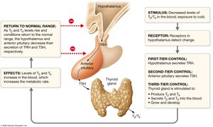
Controlled by a negative feedback loop involving TRH (hypothalamus), TSH (anterior pituitary), and thyroid hormones.
TSH stimulates production and secretion of T3 and T4, and growth of the thyroid gland.
Thyroid Disorders
Hyperthyroidism: Overproduction of thyroid hormones; causes weight loss, heat intolerance, heart rhythm disruptions.
Graves Disease: Autoimmune hyperthyroidism; may cause exophthalmos and goiter.
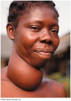
Hypothyroidism: Underproduction; causes weight gain, cold intolerance, slow heart rate, goiter.
Parathyroid Hormone and Calcitonin: Bone Homeostasis
Parathyroid Hormone (PTH): Increases blood calcium by stimulating osteoclasts, increasing intestinal absorption, and kidney reabsorption.
Calcitonin: Produced by thyroid parafollicular cells; inhibits osteoclasts, lowers blood calcium.
Structure of the Adrenal Glands
Adrenal Cortex: Three zones producing mineralocorticoids (aldosterone), glucocorticoids (cortisol), and androgenic steroids.
Adrenal Medulla: Neuroendocrine organ producing catecholamines (epinephrine, norepinephrine).
Hormones of the Adrenal Cortex
Aldosterone: Regulates sodium, potassium, fluid volume, blood pressure, and acid-base balance.
Cortisol: Mediates stress response, increases gluconeogenesis, anti-inflammatory effects.
Androgenic Steroids: Affect reproductive organs and other tissues.
Hormones of the Adrenal Medulla
Chromaffin Cells: Secrete epinephrine and norepinephrine; mediate immediate stress response.
Effects: Increase heart rate, dilate bronchioles, constrict/dilate blood vessels, decrease digestive/urinary functions.
Structure of the Pancreas
Pancreatic Islets: Endocrine cells (alpha, beta, delta) secrete glucagon, insulin, and somatostatin.
Acinar Cells: Exocrine cells secrete digestive enzymes.
Hormones of the Endocrine Pancreas: Glucose Homeostasis
Glucagon: Increases blood glucose by promoting glycogenolysis, gluconeogenesis, protein/fat breakdown, ketone body formation.
Insulin: Lowers blood glucose by promoting uptake and storage of nutrients; required for glucose uptake by target cells.
Diabetes Mellitus: Type 1 (autoimmune destruction of beta cells), Type 2 (insulin resistance).
Blood Glucose Regulation
Negative feedback loops regulate blood glucose via insulin and glucagon secretion.
The Thymus: Thymosin and Thymopoietin
Site of T lymphocyte maturation; secretes hormones assisting immune development.
The Gonads: Sex Hormones
Testes: Produce testosterone (anabolic and androgenic effects).
Ovaries: Produce estrogens and progesterone (secondary sex characteristics, menstrual cycle, pregnancy support).
The Pineal Gland: Melatonin
Secretes melatonin; regulates sleep-wake cycles.
Adipose Tissue: Leptin
Leptin induces satiety; amount correlates with adipose tissue.
The Heart: Atrial Natriuretic Peptide
ANP causes vasodilation and enhances sodium/water excretion, lowering blood pressure.
The Kidneys: Erythropoietin, Renin, Vitamin D Activation
EPO stimulates erythrocyte production; renin activates angiotensin system; kidneys convert vitamin D to active form.
Hormonal Control of Metabolic and Fluid Homeostasis
Thyroid hormones set basal metabolic rate; insulin and glucagon regulate glucose during feeding/fasting; catecholamines and glucagon increase during exercise.
ADH, aldosterone, and ANP regulate fluid balance and urine output.
Summary Table: Hormones, Stimuli, Target Tissues, and Effects
Hormone | Stimulus | Target Tissue | Effect |
|---|---|---|---|
ADH | High solute concentration | Kidneys | Water reabsorption |
Oxytocin | Uterine stretch, suckling | Uterus, mammary glands | Contractions, milk ejection |
TSH | TRH | Thyroid gland | Thyroid hormone secretion |
ACTH | CRH | Adrenal cortex | Cortisol, aldosterone secretion |
GH | GHRH, fasting, exercise | Liver, muscle, bone | Growth, protein synthesis |
Insulin | High blood glucose | Most cells | Glucose uptake |
Glucagon | Low blood glucose | Liver, muscle, adipose | Increase blood glucose |
PTH | Low blood calcium | Bone, kidney, intestine | Increase blood calcium |
Calcitonin | High blood calcium | Bone | Decrease blood calcium |
Aldosterone | High K+, low Na+, angiotensin | Kidney | Na+ retention, K+ excretion |
Cortisol | Stress, ACTH | Most tissues | Increase glucose, anti-inflammatory |
Key Equations
cAMP Formation:
Signal Amplification:
Additional info:
Some details and examples were inferred for completeness and clarity.
