 Indietro
Study Notes: The Lymphatic and Respiratory Systems
Guida di studio - Note intelligenti
 Appunti personalizzati basati sui tuoi materiali, ampliati con definizioni chiave, esempi e contesto.
The Lymphatic System and Body Defenses
Lymphatic Capillaries and Organs
The lymphatic system is a crucial component of the body's immune defenses, consisting of a network of vessels and organs that help maintain fluid balance and protect against pathogens.
Lymphatic capillaries are more permeable than blood capillaries, allowing them to absorb larger molecules and particles.
Other lymphoid organs include the spleen, thymus, tonsils, Peyer's patches, and appendix. These organs contribute to immune function by housing and supporting lymphocytes.
Lymphoid tissue is found in various submucosal membrane sites throughout the body, such as the gastrointestinal tract, oral passage, nasopharyngeal tract, thyroid, breast, lung, salivary gland, eye, and skin.
Lymphoid tissues are populated by lymphocytes, macrophages, and plasma cells.
Mucosa-Associated Lymphatic Tissue (MALT)
MALT is a diffuse system of small concentrations of lymphoid tissue found in mucosal membranes throughout the body. It plays a key role in immune surveillance and response.
MALT includes structures such as tonsils, Peyer's patches, and lymphoid nodules in the gastrointestinal and respiratory tracts.
Categories of Immunity
The immune system is divided into two main categories: innate (nonspecific) and adaptive (specific) immunity.
Innate Immunity: Provides nonspecific resistance through physical barriers (skin, tears, saliva, mucous membranes, mucus). The response is the same every time and acts rapidly.
Adaptive Immunity: Provides specific resistance, characterized by specificity (recognizing particular substances) and memory (responding more rapidly and strongly to previously encountered substances).
Adaptive Immunity: Antigens and Self-Antigens
Adaptive immunity involves the recognition, response, and memory of specific antigens.
Antigens: Large molecules, usually foreign, that stimulate an immune response. Examples include bacteria, viruses, pollen, animal dander, foods, and drugs.
Self-antigens: Molecules produced by the body used as markers to differentiate self from non-self. Response to self-tumor antigens is helpful, but response to other self-antigens can result in autoimmune diseases.
Cellular and Antibody-Mediated Immune Responses
The adaptive immune response is divided into two arms: antibody-mediated and cell-mediated immunity.
B cells secrete antibodies and protect against extracellular antigens (bacteria, toxins, parasites, viruses outside cells).
T cells fight antigens directly and protect against intracellular antigens (viruses, intracellular bacteria, fungi, tumors).
Helper T cells and regulatory T cells regulate both arms of the adaptive immune response.
Origin and Development of Lymphocytes
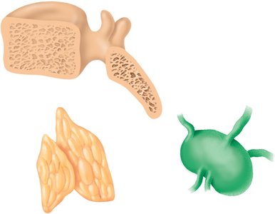
B and T lymphocytes originate in the red bone marrow and mature in primary lymphatic organs.
B cells mature in the bone marrow.
T cells mature in the thymus.
In secondary lymphatic organs and tissues (diffuse lymphatic tissue, lymphatic nodules, tonsils, lymph nodes, spleen), lymphocytes interact with antigens and antigen-presenting cells to produce the immune response.
Activation of Lymphocytes
For an effective immune response, lymphocytes must recognize antigens and proliferate to destroy them.
Lymphocytes recognize the antigen.
After recognition, lymphocytes increase in number to effectively destroy the antigen.
The Respiratory System
Functional Anatomy of the Respiratory System
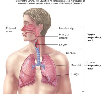
The respiratory system is responsible for gas exchange, supplying oxygen to the body and removing carbon dioxide.
Organs include the nose, pharynx, larynx, trachea, bronchi, and lungs (alveoli).
Movement of Oxygen and Carbon Dioxide
Respiration involves the movement of oxygen from the external environment to cells and the removal of carbon dioxide produced by cells.
External respiration: Exchange of oxygen and carbon dioxide in the lungs.
Internal respiration: Exchange of oxygen and carbon dioxide in the tissues.
Cellular respiration: Cells utilize oxygen and produce carbon dioxide.
Anatomy of the Upper Respiratory Tract
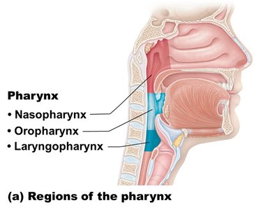
The upper respiratory tract includes the nasal cavity, pharynx, and larynx, which filter, warm, and moisten incoming air.
The nose contains conchae, which increase surface area and air turbulence, aiding in trapping inhaled particles.
The palate separates the nasal cavity from the oral cavity (hard palate anterior, soft palate posterior).
The Larynx and Trachea
The larynx (voice box) routes air and food into proper channels and plays a role in speech. The trachea (windpipe) connects the larynx to the bronchi and is reinforced with C-shaped rings of hyaline cartilage.
The trachea is lined with ciliated mucosa; cilia beat in a superior direction, and goblet cells produce mucus.
Lungs and Thoracic Cavity
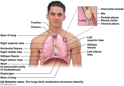
The lungs are divided into lobes (left lung: 2 lobes; right lung: 3 lobes) and are housed within the thoracic cavity, which is separated from the abdominal cavity by the diaphragm.
Respiratory Zone Structures
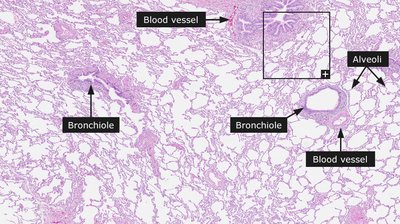
The respiratory zone is the site of gas exchange and includes respiratory bronchioles, alveolar ducts, alveolar sacs, and alveoli.
Conducting zone structures include all other passageways that transport air but do not participate in gas exchange.
Mechanics of Breathing
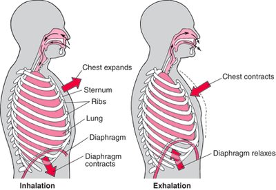
Pulmonary ventilation consists of two phases: inspiration and expiration.
Inspiration: Diaphragm and external intercostal muscles contract, increasing intrapulmonary volume and decreasing gas pressure, causing air to flow into the lungs.
Expiration: Passive process relying on lung elasticity; intrapulmonary volume decreases, gas pressure increases, and air flows out.
Forced expiration involves contraction of internal intercostal muscles and abdominal muscles.
Blood Supply to the Lungs
The lungs receive both oxygenated and deoxygenated blood.
Bronchial arteries supply oxygenated blood to lung tissues.
Pulmonary arteries bring deoxygenated blood to the lungs for oxygenation; blood leaves via pulmonary veins.
Ventilation and Boyle's Law
Ventilation is the movement of air into and out of the lungs, governed by Boyle's Law.
Air moves from areas of higher pressure to lower pressure.
Boyle's Law: (pressure is inversely proportional to volume).
As alveolar volume increases (diaphragm contracts), pressure decreases and air flows in.
Pleura and Pleural Pressure
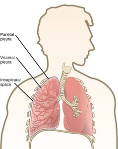
The lungs are surrounded by a double-layered membrane called the pleura.
Visceral pleura: Covers the lungs.
Parietal pleura: Lines the thoracic cavity.
Pleural pressure is negative, helping alveoli expand.
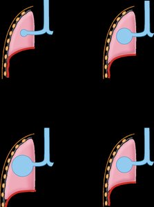
Pneumothorax is a condition where an opening between the pleural cavity and air causes loss of pleural pressure.
Intra-Alveolar Pressure Changes
Changes in intra-alveolar pressure drive the movement of air during breathing.
During inspiration, increased thoracic volume leads to decreased intra-alveolar pressure, and air moves into the lungs.
During expiration, decreased thoracic volume leads to increased intra-alveolar pressure, and air moves out of the lungs.
The Respiratory Membrane
The respiratory membrane is the site of gas exchange between alveoli and capillaries.
Three types of cells: Type I pneumocytes (gas exchange), Type II pneumocytes (produce surfactant), and dust cells (phagocytes).
Surfactant reduces surface tension, preventing alveolar collapse.
Respiratory distress syndrome occurs when insufficient surfactant is produced, common in premature infants.
Compliance and Regulation of Ventilation
Compliance measures the ease with which the lungs and thorax expand. Regulation of ventilation involves both voluntary and involuntary control.
Lower compliance means harder expansion (e.g., pulmonary fibrosis, edema, asthma).
Respiration can be modified by emotions and conscious control.
Apnea is cessation of breathing; hyperventilation is rapid breathing that decreases blood CO2.
Chemical Control of Ventilation
Chemoreceptors respond to changes in blood pH, CO2, and O2 levels to regulate breathing.
Medullary chemoreceptors respond to CO2 and pH changes.
Carotid and aortic bodies respond to pH and O2 changes.
Hypercapnia: high CO2; hypocapnia: low CO2; hypoxia: low O2.
Physical Principles of Gas Exchange
Gas exchange across the respiratory membrane depends on membrane thickness, diffusion coefficient, surface area, and partial pressure differences.
Gas moves from areas of higher partial pressure to lower partial pressure.
Diseases like emphysema and lung cancer reduce surface area for gas exchange.
Regulation of Blood pH and Gases
The respiratory system helps regulate blood pH by controlling CO2 levels.
Increased breathing decreases blood CO2 and increases pH.
Decreased breathing increases blood CO2 and decreases pH.
Summary Table: Types of Cells in the Respiratory Membrane
Cell Type | Main Function |
|---|---|
Type I pneumocyte | Gas exchange |
Type II pneumocyte | Produce surfactant |
Dust cell (phagocyte) | Remove debris and pathogens |
Summary Table: Categories of Immunity
Category | Key Features |
|---|---|
Innate Immunity | Nonspecific, rapid, physical barriers |
Adaptive Immunity | Specific, memory, B and T cells |
