 Indietro
Study Notes: The Respiratory System (Anatomy & Physiology)
Guida di studio - Note intelligenti
 Appunti personalizzati basati sui tuoi materiali, ampliati con definizioni chiave, esempi e contesto.
The Respiratory System
Overview and Functions
The respiratory system is responsible for the movement of air and the exchange of gases between the body and the environment. It consists of structures that facilitate breathing, protect respiratory surfaces, produce sounds, and aid in the sense of smell.
Gas Exchange: Provides a surface for the exchange of oxygen and carbon dioxide.
Air Movement: Moves air to and from the lungs.
Protection: Protects respiratory surfaces from pathogens and debris.
Sound Production: The larynx produces sounds for speech.
Olfaction: Assists in the sense of smell.
Anatomy of the Respiratory System
The respiratory system is divided into the upper and lower respiratory tracts.
Upper Respiratory System: Includes the nose, nasal cavity, paranasal sinuses, and pharynx.
Lower Respiratory System: Includes the larynx, trachea, bronchi, and lungs.
Pleural Cavities and Lungs
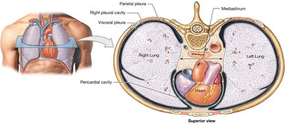
Each lung is surrounded by a pleural cavity lined by two pleural membranes:
Parietal Pleura: Lines the inside of the pleural cavity.
Visceral Pleura: Covers the surface of the lungs.
Pleural Fluid: Lubricates the space between the pleurae, reducing friction during breathing.
The Path of Air
Air enters through the nostrils, passes through the nasal cavity (where it is warmed and filtered), and moves through the pharynx, larynx, trachea, and bronchi before reaching the lungs.
Nasal Conchae: Increase surface area and create turbulence for warming air.
Pharynx: Divided into nasopharynx, oropharynx, and laryngopharynx; serves both respiratory and digestive functions.
Larynx: Site of sound production.
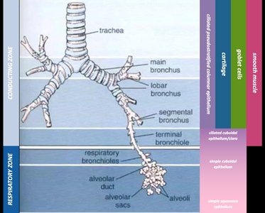
Trachea: Carries air to the bronchi; lined with ciliated epithelium to trap particles.
Bronchi and Bronchioles: Branch into smaller airways, ending in terminal and respiratory bronchioles.
Conducting and Respiratory Zones
The respiratory tract is divided into two functional zones:
Conducting Zone: Trachea, bronchi, and bronchioles; moves air but does not participate in gas exchange.
Respiratory Zone: Respiratory bronchioles, alveolar ducts, alveolar sacs, and alveoli; site of gas exchange.
Alveolar Structure and Gas Exchange
Alveoli are tiny air sacs where gas exchange occurs.
Type I Pneumocytes: Simple squamous epithelial cells; form the thin wall of the alveolus for rapid diffusion.
Type II Pneumocytes: Produce surfactant, which reduces surface tension and prevents alveolar collapse.
Alveolar Macrophages: Remove debris and pathogens from the alveolar space.
Blood-Air Barrier: Consists of the alveolar cell layer, capillary endothelial layer, and fused basement membrane; allows rapid diffusion of gases.
Respiratory Diseases and Conditions
Pneumothorax: Air enters the pleural space, causing lung collapse (atelectasis).
Respiratory Distress Syndrome: Insufficient surfactant production; increased effort to keep alveoli open.
Emphysema: Destruction of alveolar surfaces, often due to smoking.
Pneumonia: Inflammation causes fluid leakage into alveoli, restricting airflow.
Pulmonary Embolism: Blockage of pulmonary arteries.
Mechanics of Pulmonary Ventilation
Pulmonary ventilation is the physical movement of air in and out of the lungs, governed by Boyle’s law:
Boyle’s Law: (Pressure is inversely proportional to volume)
Inhalation: Thoracic cavity expands, pressure decreases, air flows in.
Exhalation: Thoracic cavity contracts, pressure increases, air flows out.
Respiratory Muscles
Primary Muscles: Diaphragm and external intercostals.
Accessory Muscles: Sternocleidomastoid, scalenes, pectoralis minor, serratus anterior, transversus thoracis, internal intercostals, rectus abdominis.
Modes of Breathing
Quiet Breathing: Inhalation is active, exhalation is passive; only primary muscles are used.
Forced Breathing: Both inhalation and exhalation are active; accessory muscles are involved.
Lung Volumes and Capacities
Tidal Volume (TV): Amount of air moved in one cycle.
Expiratory Reserve Volume (ERV): Air forcibly exhaled beyond TV.
Inspiratory Reserve Volume (IRV): Air forcibly inhaled beyond TV.
Residual Volume: Air remaining after maximum exhalation.
Vital Capacity:
Gas Diffusion and Exchange
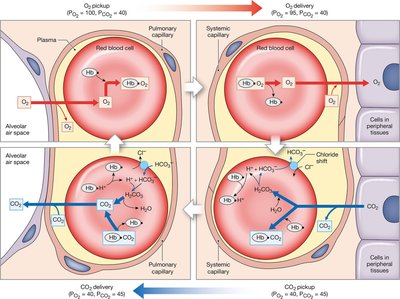
Gas exchange occurs by diffusion, driven by partial pressures of gases.
Partial Pressure: The pressure exerted by a single gas in a mixture.
External Respiration: Exchange of O2 and CO2 between alveoli and blood.
Internal Respiration: Exchange of O2 and CO2 between blood and tissues.
Oxygen and Carbon Dioxide Transport
Oxygen: Bound to hemoglobin in red blood cells.
Carbon Dioxide:
7% dissolved in plasma
23% bound to hemoglobin (carbaminohemoglobin)
70% as bicarbonate ions (HCO3-)
Enzyme: Carbonic anhydrase catalyzes this reaction.
Chloride Shift: Chloride ions move into RBCs as bicarbonate ions exit.
Regulation of Respiration
Local Control: Changes in tissue metabolism alter local partial pressures, affecting bronchodilation and bronchoconstriction.
Neural Control: Respiratory centers in the medulla and pons regulate rate and depth of breathing; reciprocal inhibition between inspiratory and expiratory neurons.
Reflex Control:
Inflation reflex prevents overexpansion.
Deflation reflex stimulates inspiration when lungs deflate.
Chemoreceptors respond to pH and CO2 levels in cerebrospinal fluid.
Review Questions
What kind of epithelium is found in the trachea? Ciliated pseudostratified columnar epithelium; traps and moves particles.
What kind of epithelium is found in the alveoli? Simple squamous epithelium; allows rapid gas diffusion.
What is the function of type II pneumocytes? Produce surfactant to reduce surface tension and keep alveoli open.
How might cigarette smoking affect breathing functions? Destroys alveolar surfaces (emphysema), increases risk of respiratory diseases.
What is the difference between quiet and forced breathing? Quiet breathing uses only primary muscles and passive exhalation; forced breathing uses accessory muscles and active exhalation.
What is meant by expiratory reserve volume? Amount of air forcibly exhaled beyond tidal volume.
Is PO2 higher or lower in the tissues (compared to capillaries)? Lower in tissues, so O2 leaves capillaries and enters tissues.
What is the enzyme involved in making bicarbonate ions? Carbonic anhydrase.
Additional info: These notes expand on lecture points with definitions, examples, and equations for clarity and completeness.
