 Indietro
Synapses and Graded Potentials: Structure and Function in Neuronal Communication
Guida di studio - Note intelligenti
 Appunti personalizzati basati sui tuoi materiali, ampliati con definizioni chiave, esempi e contesto.
Neuronal Synapses
Types and Structure of Synapses
Synapses are specialized junctions where signals are transmitted between neurons or between a neuron and its target cell, such as a muscle fiber. Neuronal synapses specifically refer to connections between two neurons, and they are essential for the integration and propagation of information in the nervous system.
Axodendritic synapse: Connection between an axon and a dendrite.
Axosomatic synapse: Connection between an axon and a cell body.
Axoaxonic synapse: Connection between an axon and another axon.
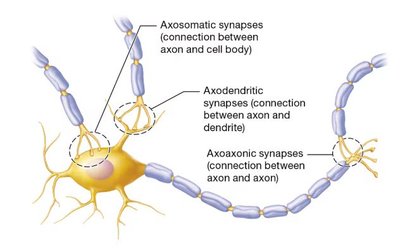
Presynaptic neuron: The neuron sending the signal from its axon terminal.
Postsynaptic neuron: The neuron receiving the signal, which may be at its dendrite, cell body, or axon.
Synaptic transmission: The process of transferring chemical or electrical signals across a synapse.
Benefits of Neuronal Synapses
Neuronal synapses provide several critical functions:
Voluntary movement
Cognition
Sensation
Emotion
Types of Synapses
Electrical Synapses
Electrical synapses occur between cells that are electrically coupled via gap junctions. These allow ions to flow directly from one neuron to another, enabling rapid and synchronized transmission.
Bidirectional: Signals can travel in both directions.
Instantaneous: Transmission occurs almost immediately.
Location: Found in regions responsible for automatic cyclic behaviors (e.g., breathing), cardiac muscle, and visceral smooth muscle.
Chemical Synapses
Most synapses in the nervous system are chemical. Here, the electrical signal is converted into a chemical signal via neurotransmitters, which then reconvert to an electrical signal in the postsynaptic neuron.
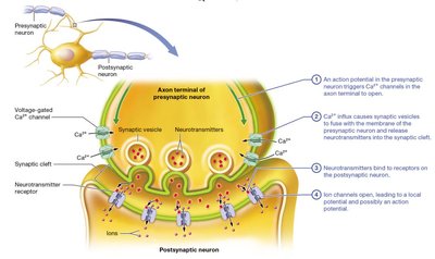
Synaptic vesicles: Contain neurotransmitters in the presynaptic axon terminal.
Synaptic cleft: The small gap filled with extracellular fluid and proteins between the presynaptic and postsynaptic neurons.
Neurotransmitter receptors: Located on the postsynaptic neuron; neurotransmitters must bind to these to transmit the signal.
Synaptic delay: Chemical synapses transmit signals more slowly than electrical synapses due to the time required for neurotransmitter release and binding.
Comparison of Electrical and Chemical Synapses
Speed: Electrical synapses are faster and bidirectional; chemical synapses are slower and unidirectional.
Signal variation: Chemical synapses allow for signal modulation and integration.
Events at a Chemical Synapse
Stepwise Process
An action potential in the presynaptic neuron triggers voltage-gated calcium ion channels to open.
Calcium influx causes synaptic vesicles to fuse with the presynaptic membrane and release neurotransmitters into the synaptic cleft.
Neurotransmitters bind to receptors on the postsynaptic neuron.
Ion channels open, leading to a local potential and possibly an action potential in the postsynaptic neuron.
Postsynaptic Potentials
Excitatory and Inhibitory Postsynaptic Potentials (EPSPs and IPSPs)
Postsynaptic potentials are local changes in membrane potential in the postsynaptic neuron.
EPSP (Excitatory Postsynaptic Potential): Small local depolarization that moves the membrane potential closer to threshold, increasing the likelihood of an action potential.
IPSP (Inhibitory Postsynaptic Potential): Small local hyperpolarization that moves the membrane potential farther from threshold, decreasing the likelihood of an action potential.
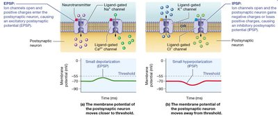
Mechanisms of EPSP and IPSP
EPSP: Occurs when ligand-gated channels open and positively charged ions (e.g., Na+, Ca2+) enter the postsynaptic neuron.
IPSP: Occurs when ligand-gated K+ channels open (cytosol loses positive charge) or ligand-gated Cl- channels open (Cl- enters cytosol), making the membrane more negative.
Neural Integration and Summation
Integration of Postsynaptic Potentials
Neurons typically receive input from multiple presynaptic sources, resulting in a combination of EPSPs and IPSPs. The cumulative effect determines whether the neuron will fire an action potential.
Neural integration: The process of combining excitatory and inhibitory inputs.
Summation: The additive effect of multiple postsynaptic potentials.
Types of Summation
Temporal summation: Rapid, repeated release of neurotransmitters from a single presynaptic neuron. Successive EPSPs bring the trigger zone to threshold.
Spatial summation: Simultaneous release of neurotransmitters from multiple presynaptic neurons. Large quantities of neurotransmitters are released at once, raising the membrane potential to threshold.
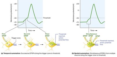
Both EPSPs and IPSPs can summate; the net effect depends on which is stronger.
Termination of Synaptic Transmission
Mechanisms of Termination
Synaptic transmission must be terminated before the postsynaptic neuron can respond again. This occurs by three main mechanisms:
Diffusion and absorption: Neurotransmitters diffuse away from the synaptic cleft and are absorbed by neurons or astrocytes.
Degradation: Enzymatic breakdown of neurotransmitters in the synaptic cleft.
Reuptake: Neurotransmitters are transported back into the presynaptic neuron by specific proteins.
Clinical Application: Neurotoxins and Synaptic Function
Effects of Arthropod Venom
Certain arthropod venoms contain neurotoxins that disrupt synaptic transmission:
Black widow spider: Causes massive release of neurotransmitters, leading to repetitive stimulation of postsynaptic neurons.
Bark scorpion: Prevents inactivation of voltage-gated sodium channels, causing continuous firing of action potentials.
Symptoms: Muscle hyperexcitability, cramps, sweating, nausea, vomiting, difficulty breathing.
Key Terms and Definitions
Synapse: Junction between two neurons or a neuron and its target cell.
Presynaptic neuron: Neuron sending the signal.
Postsynaptic neuron: Neuron receiving the signal.
Synaptic transmission: Transfer of signals across a synapse.
Electrical synapse: Direct ion flow via gap junctions.
Chemical synapse: Signal transmission via neurotransmitters.
Synaptic vesicle: Membrane-bound container for neurotransmitters.
Neurotransmitter: Chemical messenger released at synapses.
Synaptic cleft: Gap between presynaptic and postsynaptic neurons.
Neurotransmitter receptor: Protein on postsynaptic neuron that binds neurotransmitter.
EPSP: Excitatory postsynaptic potential.
IPSP: Inhibitory postsynaptic potential.
Neural integration: Summation of postsynaptic potentials.
Temporal summation: Rapid, repeated input from one neuron.
Spatial summation: Simultaneous input from multiple neurons.
Relevant Equations
Membrane Potential Change
Membrane potential changes can be described by:
Where:
\Delta V: Change in membrane potential
I: Current (ion flow)
R: Resistance of the membrane
Threshold for Action Potential
The threshold is the membrane potential at which voltage-gated channels open to initiate an action potential:
Typical resting membrane potential:
EPSPs and IPSPs shift the membrane potential toward or away from threshold, respectively.
Additional info: Summation and integration of postsynaptic potentials are fundamental for neural computation and information processing in the central nervous system.
