 Indietro
The Appendicular Skeleton: Structure and Function
Guida di studio - Note intelligenti
 Appunti personalizzati basati sui tuoi materiali, ampliati con definizioni chiave, esempi e contesto.
The Appendicular Skeleton
Overview of the Appendicular Skeleton
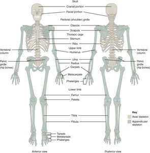
The appendicular skeleton consists of 126 bones that form the limbs and the pectoral and pelvic girdles. These structures are essential for movement and interaction with the environment, providing attachment points for muscles and facilitating articulation with the axial skeleton.
Pectoral (shoulder) girdle: Connects the upper limbs to the trunk.
Pelvic girdle: Connects the lower limbs to the trunk.
Upper and lower limbs: Enable a wide range of movements.
The Pectoral Girdle
Structure and Articulations
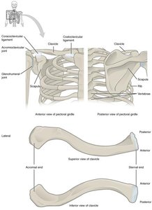
The pectoral girdle is composed of the clavicles and scapulae, providing the only bony attachment of the upper limb to the axial skeleton. It allows for a high degree of mobility at the shoulder joint.
Clavicle (collarbone): S-shaped bone that articulates medially with the manubrium of the sternum (sternoclavicular joint) and laterally with the acromion of the scapula (acromioclavicular joint).
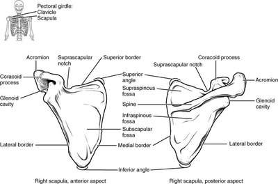
Scapula (shoulder blade): Broad, flat, triangular bone with a glenoid cavity that articulates with the humerus to form the glenohumeral (shoulder) joint.
Bones of the Upper Limb
Regions and Major Bones
The upper limb is divided into the arm (brachium), forearm (antebrachium), and hand. Each upper limb contains 30 bones, allowing for complex movements and dexterity.
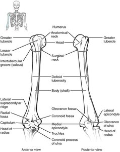
Arm (Brachium): Contains the humerus, which articulates proximally with the scapula and distally with the radius and ulna.
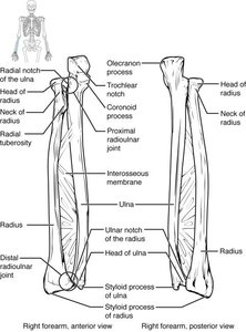
Forearm (Antebrachium): Composed of the radius (lateral) and ulna (medial).
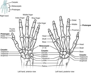
Hand: Includes the carpal bones (wrist), metacarpals (palm), and phalanges (fingers).
Key Articulations and Features
Humerus: Proximal head articulates with the glenoid cavity; distal end forms the elbow joint with the radius and ulna.
Ulna: Medial bone of the forearm; olecranon forms the point of the elbow; trochlear notch articulates with the humerus.
Radius: Lateral bone of the forearm; head articulates with the capitulum of the humerus and the radial notch of the ulna.
Carpal bones: Eight bones in two rows (proximal and distal) form the wrist.
Metacarpals: Five bones numbered I–V from thumb to little finger.
Phalanges: Fourteen bones in each hand; thumb (pollex) has two, fingers have three each.
The Pelvic Girdle and Pelvis
Structure and Function
The pelvic girdle consists of two coxal (hip) bones, each formed by the fusion of the ilium, ischium, and pubis. The pelvis supports the weight of the upper body and protects pelvic organs.
Ilium: Superior and largest part of the coxal bone.
Ischium: Posteroinferior part; ischial tuberosity bears body weight when sitting.
Pubis: Anterior part; joined at the pubic symphysis.
Acetabulum: Deep socket for articulation with the femoral head (hip joint).
Obturator foramen: Large opening covered by connective tissue for muscle attachment.
Sacroiliac joint: Articulation between the ilium and sacrum, connecting the pelvic girdle to the axial skeleton.
Bones of the Lower Limb
Regions and Major Bones
The lower limb is composed of 30 bones, including the femur, patella, tibia, fibula, tarsals, metatarsals, and phalanges. These bones support body weight and enable locomotion.
Femur: Longest and heaviest bone; articulates with the acetabulum proximally and tibia distally.
Patella: Sesamoid bone embedded in the quadriceps tendon.
Tibia: Medial, weight-bearing bone of the leg; forms the knee and ankle joints.
Fibula: Lateral, slender bone; mainly for muscle attachment.
Tarsal bones: Seven bones form the ankle, including the talus (articulates with tibia and fibula) and calcaneus (heel bone).
Metatarsals: Five bones numbered I–V from medial to lateral.
Phalanges: Fourteen bones in each foot; hallux (great toe) has two, other toes have three each.
Summary Table: Major Bones of the Appendicular Skeleton
Region | Main Bones | Key Joints |
|---|---|---|
Pectoral Girdle | Clavicle, Scapula | Sternoclavicular, Acromioclavicular, Glenohumeral |
Upper Limb | Humerus, Radius, Ulna, Carpals, Metacarpals, Phalanges | Elbow, Radioulnar, Wrist |
Pelvic Girdle | Ilium, Ischium, Pubis | Sacroiliac, Pubic Symphysis, Hip |
Lower Limb | Femur, Patella, Tibia, Fibula, Tarsals, Metatarsals, Phalanges | Knee, Ankle |
