 Indietro
The Appendicular Skeleton: Structure and Function
Guida di studio - Note intelligenti
 Appunti personalizzati basati sui tuoi materiali, ampliati con definizioni chiave, esempi e contesto.
The Skeletal System: Overview
Axial vs. Appendicular Skeleton
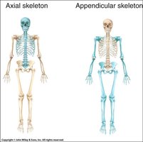
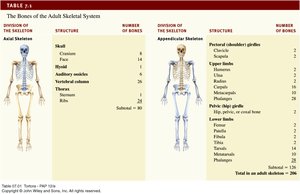
The human skeleton is divided into two major parts: the axial skeleton and the appendicular skeleton. The axial skeleton forms the central axis of the body, while the appendicular skeleton consists of the limbs and girdles that attach them to the axial skeleton.
Axial Skeleton: Includes the skull, vertebral column, and thoracic cage.
Appendicular Skeleton: Includes the pectoral girdle, pelvic girdle, and the bones of the upper and lower limbs.
The Appendicular Skeleton
Pectoral Girdle
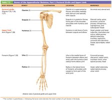
The pectoral girdle, also known as the shoulder girdle, supports the upper limbs and provides extensive mobility. It consists of two bones on each side: the clavicle and the scapula.
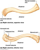
Clavicle: An elongated S-shaped bone that articulates medially with the sternum (sternoclavicular joint) and laterally with the scapula (acromioclavicular joint).
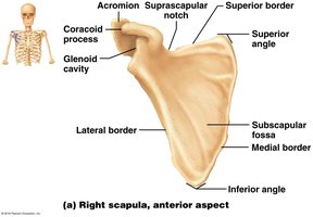
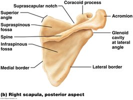
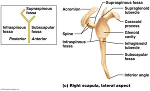
Scapula: A thin, triangular bone located on the dorsal surface of the rib cage, with three borders (superior, medial, lateral) and three angles (superior, lateral, inferior).
Upper Limb
The upper limb is divided into four regions: brachium (arm), antebrachium (forearm), carpus (wrist), and manus (hand). Each limb contains 30 bones.
Brachium: Contains the humerus, the largest bone of the upper limb.
Antebrachium: Contains the radius (lateral) and ulna (medial), which are parallel in anatomical position.
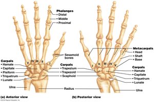
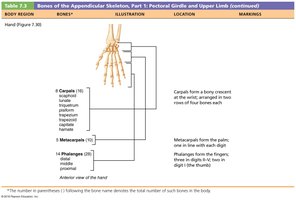
Carpus: The wrist, composed of eight carpal bones arranged in two rows.
Manus: The hand, composed of five metacarpals and fourteen phalanges.
Humerus
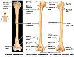
The humerus articulates with the scapula at the shoulder and with the radius and ulna at the elbow. Key features include the head, anatomical neck, greater and lesser tubercles, deltoid tuberosity, and various fossae and condyles.
Forearm: Ulna and Radius
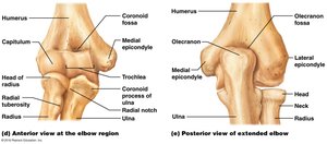
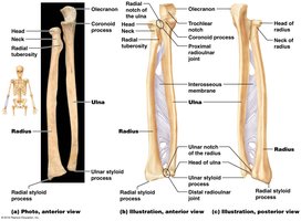
The ulna and radius are connected by the interosseous membrane and articulate with each other at both proximal and distal radioulnar joints.
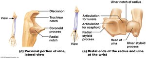
Ulna: Medial bone, forms the major portion of the elbow joint. Features include the olecranon, coronoid process, trochlear notch, and ulnar styloid process.
Radius: Lateral bone, features include the head, radial tuberosity, ulnar notch, and radial styloid process.
Hand: Carpus, Metacarpus, and Phalanges
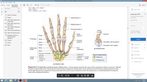
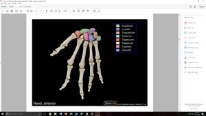
The hand consists of the carpal bones (wrist), metacarpals (palm), and phalanges (fingers). The carpal bones are arranged in two rows, and the phalanges are numbered I to V from thumb to little finger.
Pelvic Girdle and Lower Limbs
Pelvic Girdle
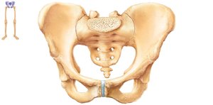
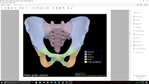
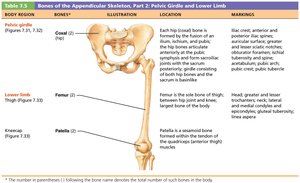
The pelvic girdle is composed of two hip bones (ossa coxae), which unite with the sacrum and coccyx to form the pelvis. The pelvis supports the trunk and protects pelvic viscera.
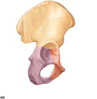
Ilium: Superior region, articulates with the sacrum.
Ischium: Posteroinferior region.
Pubis: Anterior region, joined at the pubic symphysis.
Acetabulum
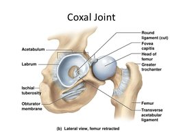
The acetabulum is the cup-shaped socket where the ilium, ischium, and pubis unite, forming the hip joint with the head of the femur.
Sexual Dimorphism of the Pelvis
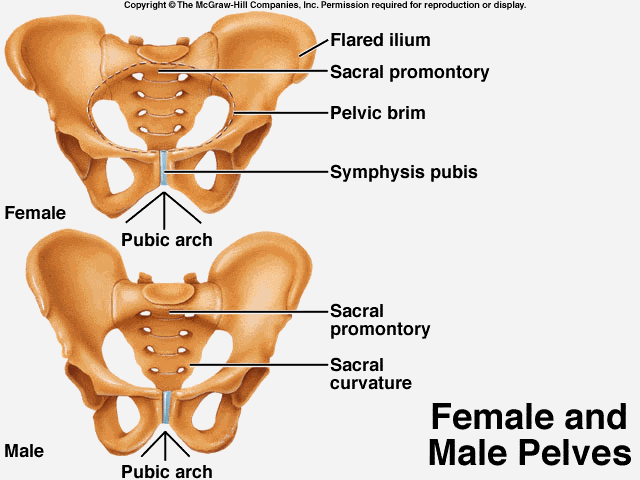
The pelvis is highly useful for determining biological sex due to differences in structure related to childbirth and locomotion. The female pelvis is wider for childbirth, while the male pelvis is optimized for bipedal locomotion.

Lower Limb
The lower limb is divided into four regions: femoral (thigh), patellar (kneecap), crural (leg), tarsal (ankle), and pedal (foot). Each limb contains 30 bones.
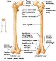
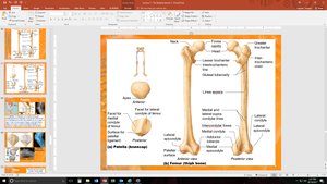
Femur: The longest and strongest bone, articulates with the acetabulum and tibia.
Patella: Sesamoid bone embedded in the tendon of the knee.
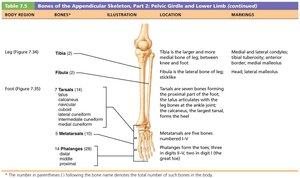
Tibia: Medial, weight-bearing bone of the leg.
Fibula: Lateral, slender bone, stabilizes the ankle.
Tarsals: Seven bones forming the ankle.
Metatarsals: Five bones forming the foot.
Phalanges: Fourteen bones forming the toes.
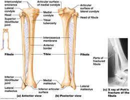
Ankle and Foot Bones
The tarsal bones are arranged in proximal and distal groups. The calcaneus forms the heel, and the talus is the most superior tarsal bone. The metatarsals and phalanges are similar in arrangement to those of the hand.
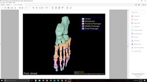
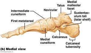
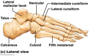
Metatarsals and Phalanges
The great toe (hallux) contains only two phalanges, while the other toes contain three. Each metatarsal and phalangeal bone has a base, body, and head.
Anatomical Checklists
Checklists are useful for reviewing the anatomical features of the pectoral girdle, upper limb, pelvic girdle, and lower limb.
Summary Table: Bones of the Appendicular Skeleton
Region | Bones | Key Features |
|---|---|---|
Pectoral Girdle | Clavicle, Scapula | Mobility, muscle attachment |
Upper Limb | Humerus, Radius, Ulna, Carpals, Metacarpals, Phalanges | Articulation, movement, grasping |
Pelvic Girdle | Ilium, Ischium, Pubis | Weight-bearing, protection |
Lower Limb | Femur, Patella, Tibia, Fibula, Tarsals, Metatarsals, Phalanges | Locomotion, support |
Additional info: The appendicular skeleton is essential for movement and interaction with the environment, providing attachment points for muscles and facilitating locomotion and manipulation.
