Indietro
The Appendicular Skeleton: Structure and Function
Guida di studio - Note intelligenti
 Appunti personalizzati basati sui tuoi materiali, ampliati con definizioni chiave, esempi e contesto.
The Appendicular Skeleton
Overview
The appendicular skeleton consists of the bones of the limbs and their girdles, which attach the limbs to the axial skeleton. It is divided into the pectoral girdle (shoulder) and pelvic girdle (hip), each providing structural support and mobility for the upper and lower limbs, respectively.
Pectoral girdle: Attaches upper limbs to the body trunk.
Pelvic girdle: Attaches lower limbs to the body trunk.
Pectoral Girdle
Structure and Function
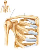
The pectoral girdle consists of the clavicles (anteriorly) and scapulae (posteriorly). It provides attachment sites for muscles that move the upper limbs and offers a high degree of mobility due to the shallow socket of the shoulder joint and the lack of direct attachment of the scapulae to the axial skeleton.
Clavicle: S-shaped bone, anchors muscles, acts as a brace to hold the scapulae and arms out laterally.
Scapula: Thin, triangular bone with three borders (superior, medial, lateral) and several important features (spine, acromion, coracoid process, glenoid cavity).
Upper Limb
Arm: Humerus
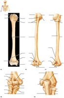
The humerus is the only bone of the arm and is the largest and longest bone of the upper limb. It articulates with the scapula at the shoulder and with the radius and ulna at the elbow.
Head: Fits into the glenoid cavity of the scapula.
Anatomical neck: Slight constriction below the head.
Greater and lesser tubercles: Sites of muscle attachment.
Surgical neck: Most frequently fractured part.
Distal features: Trochlea, capitulum, epicondyles, and fossae.
Forearm: Radius and Ulna
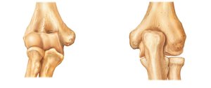
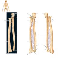
The forearm is formed by two parallel bones: the ulna (medial) and radius (lateral). They are connected by the interosseous membrane and articulate with each other both proximally and distally.
Ulna: Forms the major portion of the elbow joint with the humerus; features include olecranon, coronoid process, trochlear notch, and styloid process.
Radius: Articulates with the capitulum of the humerus and radial notch of the ulna; features include head, radial tuberosity, styloid process.
Hand: Carpus, Metacarpus, Phalanges
The hand consists of the carpus (wrist), metacarpus (palm), and phalanges (fingers). The carpus contains eight bones arranged in two rows, the metacarpus has five bones, and the phalanges are the bones of the fingers.
Carpus: Proximal row (scaphoid, lunate, triquetrum, pisiform); distal row (trapezium, trapezoid, capitate, hamate).
Metacarpals: Numbered I to V from thumb to little finger.
Phalanges: Digit I (thumb) has two bones; digits II-V have three bones each.
Pelvic Girdle
Structure and Function
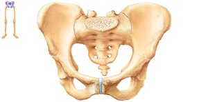
The pelvic girdle is formed by two hip bones (coxal bones or os coxae) and the sacrum. It attaches the lower limbs to the axial skeleton, transmits the weight of the upper body to the lower limbs, and supports pelvic organs. It is less mobile but more stable than the shoulder joint.
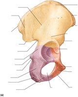
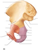
Ilium: Superior region, articulates with sacrum.
Ischium: Posteroinferior part.
Pubis: Anterior portion, joins at pubic symphysis.
Acetabulum: Deep socket for the head of the femur.
Pelvic Structure and Childbearing
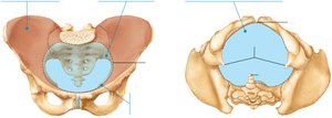
The pelvis is formed by the hip bones, sacrum, and coccyx. The female pelvis is adapted for childbearing, being wider, shallower, lighter, and rounder than the male pelvis. The pelvic brim defines the pelvic inlet, and the true pelvis defines the birth canal.
False pelvis: Superior to pelvic brim.
True pelvis: Inferior to pelvic brim.
Pelvic outlet: Inferior margin of true pelvis.
Comparison of Male and Female Pelves
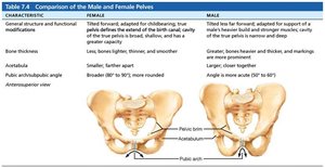
The male and female pelves differ in structure and function, reflecting adaptations for childbearing in females.
Characteristic | Female | Male |
|---|---|---|
General structure and function | Tilted forward, adapted for childbearing; true pelvis broad, shallow, and has greater capacity | Tilted less forward, adapted for support of heavier build and stronger muscles; true pelvis narrow and deep |
Bones thickness | Lighter, thinner, and smoother | Greater, bones thicker and markings more prominent |
Acetabula | Smaller, farther apart | Larger, closer together |
Pubic arch/angle | Broader (80–90°), more rounded | Angle is more acute (50–60°) |
Sacrum | Wider, shorter | Narrow, longer |
Coccyx | More movable, straighter | Less movable, curves sharply |
Lower Limb
Thigh: Femur and Patella
The femur is the largest and strongest bone in the body, articulating with the acetabulum of the hip and the tibia and patella at the knee. The patella is a sesamoid bone in the quadriceps tendon that protects the knee joint.
Femur features: Head, fovea capitis, greater and lesser trochanters, gluteal tuberosity, linea aspera, condyles, epicondyles, adductor tubercle, patellar surface, intercondylar fossa.
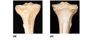
Leg: Tibia and Fibula
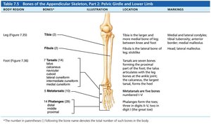
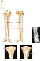
The leg is made up of two parallel bones: the tibia (medial, weight-bearing) and fibula (lateral, not weight-bearing). They are connected by the interosseous membrane and articulate with each other at both ends.
Tibia features: Medial and lateral condyles, intercondylar eminence, tibial tuberosity, anterior border, medial malleolus, fibular notch.
Fibula features: Head, lateral malleolus.
Foot
Structure
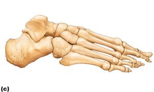
The skeleton of the foot includes the tarsus, metatarsus, and phalanges. The tarsus consists of seven bones, with the talus and calcaneus bearing most of the body weight. The metatarsals are five bones forming the foot's arch, and the phalanges are the bones of the toes.
Tarsus: Talus, calcaneus, cuboid, navicular, medial/intermediate/lateral cuneiforms.
Metatarsals: Numbered I to V from hallux (great toe) to little toe.
Phalanges: Digit I (hallux) has two bones; digits II-V have three bones each.
Copyright Notice
This work is protected by United States copyright laws and is provided solely for the use of instructors in teaching their courses and assessing student learning. Dissemination or sale of any part of this work is not permitted.