 Indietro
The Appendicular Skeleton: Structure, Function, and Development
Guida di studio - Note intelligenti
 Appunti personalizzati basati sui tuoi materiali, ampliati con definizioni chiave, esempi e contesto.
The Appendicular Skeleton
Overview
The appendicular skeleton consists of the bones of the limbs and their supporting girdles, which connect them to the axial skeleton. It is essential for movement, support, and interaction with the environment.
Pectoral girdle: Attaches upper limbs to the trunk.
Pelvic girdle: Attaches lower limbs to the trunk.
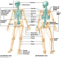
The Pectoral Girdle
Structure and Function
The pectoral girdle is composed of paired clavicles and scapulae, forming the shoulders. It provides attachment points for muscles and allows a high degree of mobility for the upper limbs.
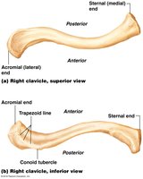
Clavicle (collarbone): S-shaped bone that acts as a brace to hold the scapula and arm laterally.
Scapula (shoulder blade): Flat, triangular bone on the dorsal surface of the rib cage.
Mobility is enhanced because the scapulae are not directly attached to the axial skeleton and the shoulder joint socket is shallow.
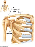
Clavicle
The clavicle articulates medially with the sternum and laterally with the scapula. It anchors muscles and acts as a strut to keep the upper limb away from the thorax.
Sternal end: Cone-shaped, articulates with the sternum.
Acromial end: Flattened, articulates with the scapula.
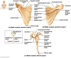
Scapula
The scapula has three borders (superior, medial, lateral) and three angles (superior, lateral, inferior). Key features include the spine, acromion, coracoid process, and several fossae for muscle attachment.
Spine: Prominent ridge on the posterior surface.
Acromion: Lateral projection forming the acromioclavicular joint.
Coracoid process: Anterior projection anchoring the biceps muscle.
Glenoid cavity: Shallow socket for the humerus.
The Upper Limb
Overview
The upper limb consists of 30 bones, divided into the arm (humerus), forearm (radius and ulna), and hand (carpals, metacarpals, phalanges).
Arm: Humerus
Forearm: Radius (lateral), Ulna (medial)
Hand: 8 carpals, 5 metacarpals, 14 phalanges
Humerus (Arm)
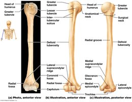
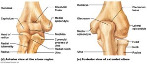
The humerus is the only bone of the arm and the largest bone of the upper limb. It articulates with the scapula proximally and the radius and ulna distally.
Head: Fits into the glenoid cavity of the scapula.
Anatomical neck: Slight constriction below the head.
Surgical neck: Common fracture site.
Greater and lesser tubercles: Muscle attachment sites.
Deltoid tuberosity: Attachment for deltoid muscle.
Trochlea and capitulum: Distal condyles for articulation with ulna and radius.
Forearm: Radius and Ulna
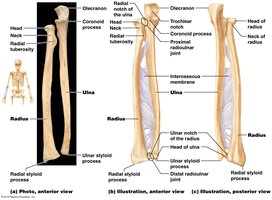
The forearm contains two parallel bones, the radius (lateral) and ulna (medial), connected by an interosseous membrane. They articulate with each other both proximally and distally.
Ulna: Forms the major portion of the elbow joint with the humerus. Features include the olecranon, coronoid process, and ulnar styloid process.
Radius: Lateral bone, articulates with the capitulum of the humerus and the radial notch of the ulna. Features include the head, radial tuberosity, and radial styloid process.
Hand: Carpals, Metacarpals, and Phalanges
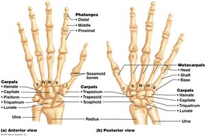
The hand consists of the carpus (wrist), metacarpus (palm), and phalanges (fingers). The carpus contains eight bones in two rows, while the metacarpus has five bones, and the phalanges are arranged in three per finger (except the thumb, which has two).
Carpal bones: Scaphoid, lunate, triquetrum, pisiform (proximal); trapezium, trapezoid, capitate, hamate (distal).
Metacarpals: Numbered I–V from thumb to little finger.
Phalanges: Proximal, middle, and distal (except thumb/pollex, which lacks a middle phalanx).
The Pelvic Girdle
Structure and Function
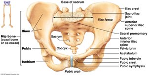
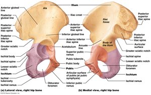
The pelvic girdle, or hip girdle, is formed by two hip bones (coxal bones) and the sacrum. It attaches the lower limbs to the axial skeleton, transmits body weight, and supports pelvic organs. It is more stable but less mobile than the pectoral girdle.
Each coxal bone is formed by the fusion of three bones: ilium, ischium, and pubis.
Acetabulum: Deep socket for the head of the femur.
Pelvic Structure and Gender Differences
The pelvis is formed by the hip bones, sacrum, and coccyx. The female pelvis is adapted for childbirth and is wider, shallower, and lighter than the male pelvis.
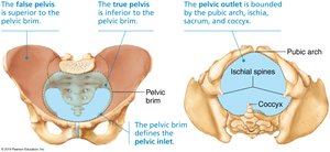
Pelvic brim (inlet): Oval ridge separating the false and true pelvis.
False pelvis: Superior to pelvic brim, supports abdominal viscera.
True pelvis: Inferior to pelvic brim, forms the birth canal.
Pelvic outlet: Inferior margin of the true pelvis.
Comparison of Male and Female Pelves
The following tables summarize the main differences between male and female pelves:
Characteristic | Female | Male |
|---|---|---|
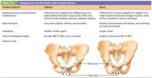
General structure | Tilted forward, adapted for childbirth, broad, shallow, greater capacity | Tilted less forward, adapted for support, narrow, deep, heavier |
Bone thickness | Lighter, thinner, smoother | Heavier, thicker, more prominent markings |
Acetabula | Smaller, farther apart | Larger, closer |
Angle of pubic arch | Broader (80–90°), more rounded | More acute (50–60°) |
Characteristic | Female | Male |
|---|---|---|
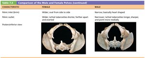
Pelvic inlet (brim) | Wider, oval | Narrow, heart-shaped |
Pelvic outlet | Wider, ischial tuberosities shorter, farther apart | Narrower, ischial tuberosities longer, sharper |
Characteristic | Female | Male |
|---|---|---|
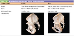
Sacrum | Wider, shorter, less curved | Narrow, longer, more curved |
Coccyx | More movable, projects inferiorly | Less movable, projects anteriorly |
Greater sciatic notch | Wide and shallow | Narrow and deep |
The Lower Limb
Overview
The lower limb supports the entire body weight and is adapted for locomotion. It consists of the thigh (femur), leg (tibia and fibula), and foot (tarsals, metatarsals, phalanges).
Thigh: Femur
Leg: Tibia (medial), Fibula (lateral)
Foot: 7 tarsals, 5 metatarsals, 14 phalanges
Femur (Thigh Bone)
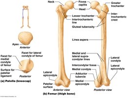
The femur is the largest and strongest bone in the body. It articulates proximally with the acetabulum and distally with the tibia and patella.
Fovea capitis: Pit for ligament attachment.
Greater and lesser trochanters: Muscle attachment sites.
Gluteal tuberosity: Attachment for gluteus maximus.
Condyles: Articulate with tibia.
Patella: Sesamoid bone protecting the knee joint.
Leg: Tibia and Fibula
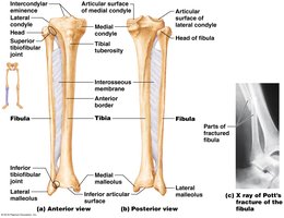
The tibia is the main weight-bearing bone of the leg, while the fibula is thinner and not weight-bearing. They are connected by an interosseous membrane.
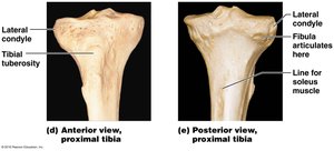
Tibia: Medial, articulates with femur and talus. Features include medial/lateral condyles, tibial tuberosity, and medial malleolus.
Fibula: Lateral, articulates with tibia. Features include head and lateral malleolus. Common site for Pott’s fracture.
Foot: Tarsals, Metatarsals, and Phalanges
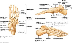
The foot is structured to support body weight and enable movement. It consists of tarsal bones (including the talus and calcaneus), metatarsals, and phalanges.
Tarsals: 7 bones, with the talus and calcaneus bearing most weight.
Metatarsals: 5 bones, numbered I–V from hallux (big toe) to little toe.
Phalanges: 14 bones, with the hallux having two and the others three each.
Arches of the Foot
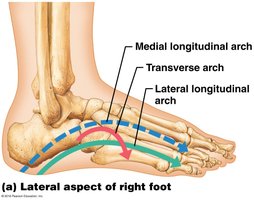
The foot has three arches (lateral longitudinal, medial longitudinal, and transverse) maintained by bones, ligaments, and tendons. These arches allow the foot to bear weight and act as a lever during movement.
Lateral longitudinal arch: Low curve on the lateral side.
Medial longitudinal arch: Higher curve on the medial side.
Transverse arch: Runs across the foot.
Developmental Aspects of the Skeleton
Infant Skull and Fontanelles
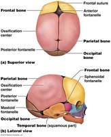
At birth, the skull has more bones than in adulthood due to unfused bones and the presence of fontanelles—fibrous membranes that allow for brain growth and ease of birth. Major fontanelles include anterior, posterior, mastoid, and sphenoidal.
Congenital Abnormalities
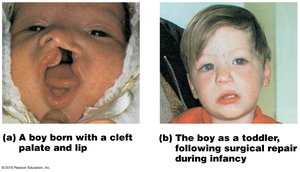
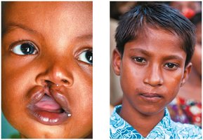
Cleft palate is a common congenital abnormality where the right and left halves of the palate fail to fuse, interfering with feeding and increasing the risk of aspiration pneumonia.
Growth and Proportions
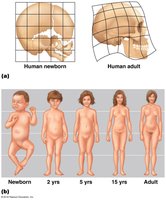
At birth, the cranium is large relative to the face. As a child grows, the arms and legs lengthen more rapidly than the head and trunk, resulting in adult body proportions.
Spinal Curvature Development
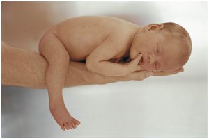
Primary spinal curvatures (thoracic and sacral) are present at birth, giving the spine a C-shape. Secondary curvatures (cervical and lumbar) develop as the child lifts its head and begins to walk.
Aging and the Skeleton
With age, intervertebral discs lose hydration and elasticity, increasing the risk of herniation and loss of stature. Costal cartilages ossify, making the thorax more rigid, and all bones lose mass.
