 Indietro
Chapter 16: The Autonomic Nervous System and Higher-Order Functions: Study Guide
Guida di studio - Note intelligenti
 Appunti personalizzati basati sui tuoi materiali, ampliati con definizioni chiave, esempi e contesto.
The Autonomic Nervous System and Higher-Order Functions
Introduction to the Autonomic Nervous System (ANS)
The autonomic nervous system (ANS) is a division of the peripheral nervous system responsible for involuntary control of visceral effectors. It operates without conscious instruction and regulates cardiovascular, respiratory, digestive, urinary, and reproductive functions. Higher-order functions include consciousness, sleep, arousal, and memory.
Somatic Nervous System (SNS): Controls voluntary movements of skeletal muscles.
Autonomic Nervous System (ANS): Controls involuntary actions of smooth muscle, cardiac muscle, glands, and adipose tissue.
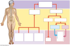
Organization of the ANS vs. SNS
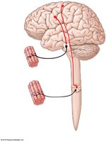
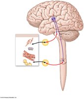
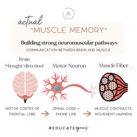
The SNS and ANS differ in their organization and function. The SNS uses a single motor neuron to innervate skeletal muscle, while the ANS uses a two-neuron chain (preganglionic and postganglionic neurons) to innervate visceral effectors.
SNS: Upper motor neurons in the primary motor cortex synapse with lower motor neurons in the brainstem or spinal cord, which directly innervate skeletal muscle.
ANS: Integrative centers in the hypothalamus send signals via preganglionic neurons to autonomic ganglia, where ganglionic neurons innervate visceral effectors.
Divisions of the ANS
The ANS consists of two main divisions: the sympathetic division and the parasympathetic division. These divisions often have opposing effects on target organs.
Sympathetic Division: Prepares the body for 'fight or flight' responses, increasing alertness, metabolic rate, and muscular abilities.
Parasympathetic Division: Promotes 'rest and digest' activities, conserving energy and maintaining resting metabolic rate.
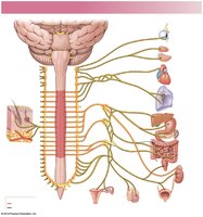
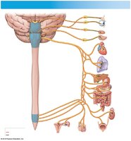
Sympathetic Division: Structure and Function
The sympathetic division is also known as the thoracolumbar division. Its preganglionic fibers are short and synapse in ganglia near the spinal cord, while postganglionic fibers are long (except at the adrenal medullae).
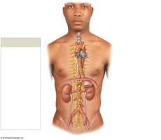
Sympathetic Chain Ganglia: Located on both sides of the vertebral column; control effectors in the body wall, thoracic cavity, head, neck, and limbs.
Collateral Ganglia: Anterior to vertebral bodies; innervate abdominopelvic tissues and viscera.
Adrenal Medullae: Modified sympathetic ganglia; release neurotransmitters/hormones directly into the bloodstream.
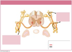
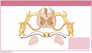
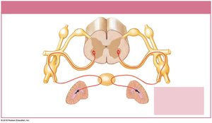
Sympathetic Neurotransmitter Release
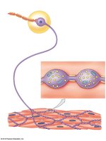
Stimulation of sympathetic preganglionic neurons releases acetylcholine (ACh) at synapses with ganglionic neurons, causing excitation. Ganglionic neurons release neurotransmitters (mostly norepinephrine, some ACh) at target organs. Varicosities are swollen segments packed with neurotransmitter vesicles.
Receptors: Target cells have membrane G protein-coupled receptors (α or β) for neurotransmitters.
Effects: Can be excitatory or inhibitory depending on receptor type.
Parasympathetic Division: Structure and Function
The parasympathetic division is also known as the craniosacral division. Its preganglionic fibers are long and originate in the brainstem and sacral segments of the spinal cord. Ganglionic neurons are located in peripheral ganglia within or adjacent to target organs, and postganglionic fibers are short.
All parasympathetic neurons release ACh.
Cholinergic Receptors: Nicotinic (excitation) and muscarinic (excitation or inhibition, longer-lasting effects).
Comparison of Sympathetic and Parasympathetic Divisions
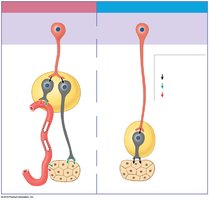
The sympathetic and parasympathetic divisions differ in neurotransmitters, ganglia locations, and effects on target organs.
Division | Preganglionic Neurotransmitter | Postganglionic Neurotransmitter | Effect |
|---|---|---|---|
Sympathetic | ACh | NE (mostly), ACh (some) | Fight or flight |
Parasympathetic | ACh | ACh | Rest and digest |
Autonomic Plexuses and Dual Innervation
Most vital organs receive dual innervation from both divisions of the ANS, allowing for opposing effects and fine control. Autonomic tone refers to the baseline activity of autonomic motor neurons, even without stimulation.
Visceral Reflexes and Regulation
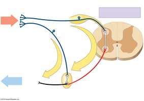
Visceral reflexes are automatic responses to stimuli involving visceral effectors. Long reflexes regulate visceral activities in most organs, while short reflexes control and coordinate the digestive tract and associated glands.
Long Reflexes: Involve CNS processing centers.
Short Reflexes: Involve the enteric nervous system.
Hierarchy of Autonomic Control
Higher levels of autonomic control are coordinated by processing centers in the medulla oblongata and regulated by the hypothalamus. These centers manage complex reflexes such as salivation, swallowing, digestive secretions, peristalsis, and urinary function.
Cranial Nerves and Reflexes
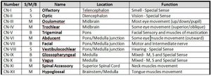
Cranial nerves are essential for sensory and motor functions, including reflexes. Monosynaptic and polysynaptic reflex arcs involving cranial nerves are clinically useful for assessing nerve and brain function.
Number | S/M/B | Name | Location | Function |
|---|---|---|---|---|
CN I | S | Olfactory | Telencephalon | Smell - Special Sense |
CN II | S | Optic | Diencephalon | Vision - Special Sense |
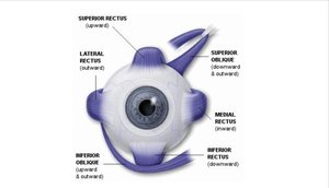
CN III | M | Oculomotor | Midbrain | Eye movement |
CN IV | M | Trochlear | Midbrain | Eye movement (superior oblique) |
CN V | B | Trigeminal | Pons | Facial sensation, mastication |
CN VI | M | Abducent | Pons/Medulla | Eye movement (lateral rectus) |
CN VII | B | Facial | Pons/Medulla | Motor and intermediate nerve |
CN VIII | S | Vestibulocochlear | Pons/Medulla | Hearing, balance |
CN IX | B | Glossopharyngeal | Medulla | Mixed, S, M, Special Sense |
CN X | B | Vagus | Medulla | Mixed, S, M, Special Sense |
CN XI | M | Spinal Accessory | Spinal Cord | Neck muscles movement |
CN XII | M | Hypoglossal | Brainstem/Medulla | Tongue muscles movement |
Somatic vs. Autonomic Function
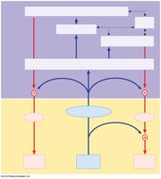
The nervous system integrates sensory information and coordinates responses through somatic and autonomic pathways. Somatic reflexes involve lower motor neurons and skeletal muscles, while autonomic reflexes involve preganglionic and ganglionic neurons affecting visceral effectors.
States of Sleep and Arousal
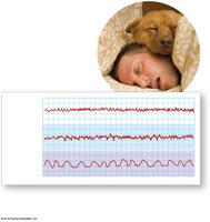
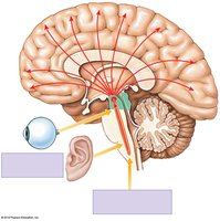
Sleep consists of REM and non-REM (deep) stages. The reticular activating system (RAS) is responsible for arousal and maintaining consciousness. EEG patterns during REM sleep resemble those of awake adults.
REM Sleep: Rapid eye movement, vivid dreams, EEG similar to awake state.
Deep Sleep: Slow wave, restorative, reduced brain activity.
Memory Formation and Storage
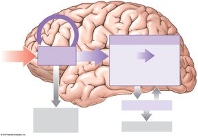
Memory is created, stored, and recalled through anatomical and physiological changes in neurons and synapses. Repetition promotes retention, converting short-term memory to long-term memory. Brain regions involved include the amygdaloid body, hippocampus, nucleus basalis, and cerebral cortex.
Short-Term Memory: Temporary, easily lost.
Long-Term Memory: Consolidated through repetition.
Cellular Mechanisms: Increased neurotransmitter release, facilitation at synapses, formation of additional synaptic connections.
Brain Chemistry and Disorders
Changes in neurotransmitter levels can affect brain function and behavior. Examples include Huntington's disease (loss of ACh- and GABA-secreting neurons), serotonin (affects mood and sensory interpretation), and dopamine (Parkinson's disease and schizophrenia).
Huntington's Disease: Difficulty controlling movements, intellectual decline.
Serotonin: Hallucinations (excess), depression/anxiety (deficiency).
Dopamine: Motor problems (deficiency), schizophrenia (excess).
Effects of Aging on the Nervous System
Aging leads to anatomical and functional changes in the nervous system, including reduction in brain size and weight, decreased number of neurons, reduced blood flow, and changes in synaptic organization. Functional changes include difficulty with memory consolidation, reduced sensory acuity, slower reaction times, weakened reflexes, and decreased motor precision. Senile dementia and Alzheimer's disease are common age-related conditions.
Integration of Nervous System with Other Body Systems
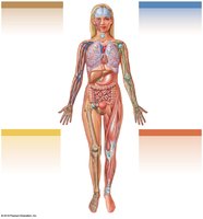
The nervous system interacts with other organ systems to maintain homeostasis and coordinate responses.
Integumentary System: Provides sensory input; nervous system controls sweat glands and arrector pili muscles.
Skeletal System: Provides calcium for neural function; nervous system affects bone maintenance.
Muscular System: Expresses emotions, communication, proprioception; nervous system controls muscle contractions.
Additional info: The notes above expand brief points into full academic explanations, add definitions, examples, and tables for clarity, and include only images that directly reinforce the educational content.
